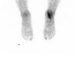
Scintigraphy, also known as a bone scan, is a medical imaging technique that uses a small amount of radioactive material to produce images of bones in the body. It is often used to evaluate the bones of the feet and to diagnose and monitor conditions such as fractures, infections, arthritis, and tumors.
During a scintigraphy procedure, a small amount of a radioactive tracer material is injected into a vein in the patient’s arm. The tracer material then travels through the bloodstream and accumulates in areas of bone that are undergoing active changes or have an increased blood supply. A special camera then captures images of the foot, showing areas of the bone that have taken up the tracer.
Scintigraphy is a safe and non-invasive procedure that typically takes between 1-3 hours to complete. Patients may be asked to lie still on a table during the procedure. The amount of radiation exposure from a bone scan is generally considered to be very low and poses little risk to the patient.
Scintigraphy for the feet can help identify bone abnormalities that may not be visible on X-rays or other imaging tests. It can also provide valuable information about the extent and severity of conditions affecting the bones of the feet, which can help guide treatment decisions.
Your doctor may recommend scintigraphy for the feet if you have symptoms such as foot pain, swelling, or limited mobility, or if they suspect a bone abnormality that cannot be identified through other imaging techniques.