Category: Radiology

MRI of your feet

Magnetic Resonance Imaging (MRI) is a medical imaging technique used to visualise internal structures of the body. It uses a strong magnetic field and radio waves to generate images of the body’s internal organs and tissues.

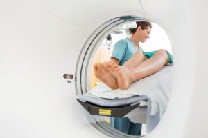
MRI can be used to diagnose a variety of foot conditions, including fractures, ligament injuries, tendon injuries, arthritis, tumours, and nerve injuries. It can also help identify soft tissue injuries that may not be visible on X-rays.

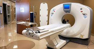
Before an MRI scan, you will be asked to remove any metal objects, including jewellery and clothing with metal zippers or buttons, as the magnetic field can cause these items to move or heat up. You may also be asked to change into a hospital gown. The MRI machine is a large, tube-shaped scanner that you will lie in. During the scan, you will need to remain still so that the images are not blurred. The procedure is painless and usually takes between 30 minutes and an hour.

After the scan, a radiologist will interpret the images and send a report to your healthcare provider. The report will include any findings and recommendations for further testing or treatment.

Computed Tomography (CT) for your feet

A CT (computed tomography) scan is a type of medical imaging test that uses X-rays and advanced computer technology to create detailed images of the bones, muscles, and other tissues in the foot. A foot CT scan can help doctors diagnose a wide range of foot and ankle conditions, such as fractures, tumours, arthritis, and deformities.

During a foot CT scan, the patient lies on a table that slides into a machine that takes multiple X-ray images of the foot from different angles. The X-ray images are then processed by a computer to create detailed cross-sectional images of the foot that can be viewed on a monitor or printed out.

Foot CT scans are typically performed on an outpatient basis and can take anywhere from 15 to 45 minutes, depending on the complexity of the scan. Before the scan, the patient may be required to remove any metal objects from their body and change into a hospital gown. After the scan, the patient can usually resume normal activities immediately. The results of the scan are typically available within a few days and will be reviewed by a doctor or radiologist.