Category: Musculoskeletal

Adult consequences of paediatric foot disorders

Paediatric foot disorders can have long-term consequences that persist into adulthood if left untreated or if not managed appropriately. Here are some of the potential adult consequences of paediatric foot disorders:

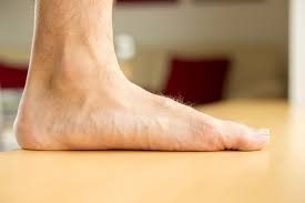
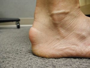
  1. Persistent foot pain: Foot disorders such as flat feet, high arches, or misaligned toes can cause pain and discomfort that can persist into adulthood.
  2. Arthritis: Certain foot conditions, such as juvenile idiopathic arthritis, can lead to the development of arthritis in adulthood.
  3. Limited mobility: If a foot disorder is left untreated or not managed appropriately, it can result in limited mobility and difficulty with activities of daily living.
  4. Balance and gait problems: Foot disorders can affect balance and gait, which can increase the risk of falls and injuries in adulthood.
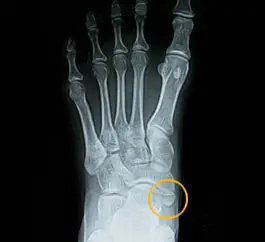
  5. Increased risk of foot and ankle injuries: Foot disorders can weaken the foot and ankle and increase the risk of injuries such as sprains, strains, and fractures.
  6. Skin and nail problems: Certain foot disorders, such as ingrown toenails or plantar warts, can persist into adulthood and cause chronic skin and nail problems.
  7. Poor self-esteem: Foot disorders can be aesthetically unappealing and may cause self-consciousness and poor self-esteem in adulthood.

It is important to address paediatric foot disorders early and to ensure that they are appropriately managed to reduce the risk of long-term consequences in adulthood. Treatment options may include orthotics, physical therapy, surgery, or a combination of these approaches. Regular follow-up with a podiatrist or foot specialist can also help to identify and address any ongoing issues.

Adult acquired flatfoot deformity (AAFD)

Adult acquired flatfoot deformity (AAFD) is a condition where the arch of the foot collapses over time, causing the foot to become flatter and wider. This can result in pain, instability, and difficulty walking or standing for long periods of time.

The most common cause of AAFD is damage to the posterior tibial tendon, which is responsible for supporting the arch of the foot. Risk factors for AAFD include age, obesity, diabetes, and prior foot or ankle injuries.

Symptoms of AAFD may include pain and swelling along the inside of the ankle, a noticeable flattening of the arch, and difficulty standing or walking on the affected foot. Some people may also experience weakness or numbness in the foot or ankle.

Treatment for AAFD may depend on the severity of the condition and the patient’s overall health. Non-surgical treatment options may include rest, ice, physical therapy, and the use of orthotic devices or supportive shoes to help alleviate pain and improve stability. In some cases, the use of braces or immobilisation devices may be necessary to reduce stress on the affected foot.

Surgical treatment for AAFD may be necessary in more severe cases, particularly if non-surgical treatments have not been successful. Surgery may involve repairing or reconstructing the damaged posterior tibial tendon, or fusing the affected joints in the foot to provide greater stability and support. Recovery from surgery may require a period of immobilisation and rehabilitation to restore strength and function to the affected foot.

Achilles non-insertional tendinopathy

Achilles non-insertional tendinopathy is a condition that affects the Achilles tendon, which connects the calf muscles to the heel bone. It is a type of tendinopathy that specifically affects the area of the tendon that is located above the heel bone, away from the point of attachment.

Symptoms of Achilles non-insertional tendinopathy may include pain and tenderness in the back of the leg, stiffness, and difficulty walking or running. The pain may worsen with activity and improve with rest.

Risk factors for Achilles non-insertional tendinopathy include overuse or repetitive strain on the Achilles tendon, improper footwear, tight calf muscles, and structural abnormalities of the foot or ankle.

Treatment for Achilles non-insertional tendinopathy may include rest, ice, physical therapy, and stretching exercises to help alleviate pain and improve flexibility. In some cases, the use of orthotic devices or supportive shoes may be recommended to reduce stress on the Achilles tendon. Non-steroidal anti-inflammatory medications may also be used to help manage pain and inflammation.

In severe cases, surgery may be necessary to repair or remove damaged tissue from the Achilles tendon. This may involve removing the damaged portion of the tendon or repairing any associated structural abnormalities in the foot or ankle. Recovery from surgery may require a period of immobilisation and rehabilitation to restore strength and function to the affected leg.

Achilles insertional tendinopathy

Achilles insertional tendinopathy is a condition that affects the Achilles tendon, which connects the calf muscles to the heel bone. It is a type of tendinopathy that specifically affects the area where the tendon attaches to the heel bone.

Symptoms of Achilles insertional tendinopathy may include pain and tenderness in the back of the heel, stiffness, and difficulty walking or running. The pain may worsen with activity and improve with rest.

Risk factors for Achilles insertional tendinopathy include overuse or repetitive strain on the Achilles tendon, improper footwear, tight calf muscles, and structural abnormalities of the foot or ankle.

Treatment for Achilles insertional tendinopathy may include rest, ice, physical therapy, and stretching exercises to help alleviate pain and improve flexibility. In some cases, the use of orthotic devices or supportive shoes may be recommended to reduce stress on the Achilles tendon. Non-steroidal anti-inflammatory medications may also be used to help manage pain and inflammation.

In severe cases, surgery may be necessary to repair or remove damaged tissue from the Achilles tendon. This may involve removing the damaged portion of the tendon and reattaching it to the heel bone or repairing any associated structural abnormalities in the foot or ankle. Recovery from surgery may require a period of immobilisation and rehabilitation to restore strength and function to the affected leg.

Achilles tendon rupture

An Achilles tendon rupture is a sudden and complete tear of the Achilles tendon, which is the large tendon that connects the calf muscles to the heel bone. This injury is most commonly seen in middle-aged men who participate in sports that involve running or jumping.

The symptoms of an Achilles tendon rupture include a sudden and severe pain in the back of the ankle, swelling, stiffness, and difficulty walking or standing on the affected leg. Some people may also hear a popping or snapping sound at the time of injury.

Treatment for an Achilles tendon rupture may depend on the severity of the injury and the patient’s overall health. Non-surgical treatment options may include rest, ice, compression, and elevation of the affected leg, as well as the use of a walking boot or cast to immobilize the ankle. Physical therapy and stretching exercises may also be recommended to help restore strength and flexibility to the ankle.

In some cases, surgical treatment may be necessary to repair the torn tendon. This may involve an open surgical procedure or a minimally invasive procedure known as percutaneous Achilles tendon repair. Recovery from an Achilles tendon rupture can take several months and may require a period of rehabilitation to regain strength and function in the affected leg.

Achilles tendon disorders

The Achilles tendon is the largest tendon in the body and connects the calf muscles to the heel bone. Achilles tendon disorders refer to a variety of conditions that can affect this tendon, causing pain and other symptoms.

Some common Achilles tendon disorders include:

  1. Achilles Tendinitis: This is an overuse injury that occurs when the tendon becomes inflamed due to repetitive stress.
  2. Achilles Tendinosis: This is a degenerative condition that occurs when the tendon is chronically overused and has micro-tears that do not heal properly.
  3. Achilles Tendon Rupture: This is a sudden and complete tear of the tendon that can occur due to a forceful contraction of the calf muscles or a traumatic injury.
  4. Haglund’s Deformity: This is a bony enlargement on the back of the heel that can irritate the Achilles tendon and cause pain.
  5. Insertional Achilles Tendinopathy: This is a condition that occurs when the Achilles tendon attaches to the heel bone and becomes inflamed or degenerated.

Symptoms of Achilles tendon disorders can include pain, swelling, stiffness, and difficulty walking or standing. Treatment for these conditions may include rest, ice, physical therapy, stretching exercises, and the use of orthotic devices or supportive shoes. In severe cases, surgery may be necessary to repair or replace the tendon.

Accessory navicular

The accessory navicular is an extra bone in the foot that is located on the inner side of the foot, near the arch. It is also known as os tibiale externum or os naviculare accessorium.

The accessory navicular bone is not present in all individuals and is considered a variation in foot anatomy. It develops during childhood and is usually fused with the navicular bone by the age of 14, although in some people it may remain separate.

In some cases, the accessory navicular can cause pain and discomfort, especially when there is excessive pressure or stress on the foot. This condition is known as accessory navicular syndrome and may cause swelling, redness, and difficulty walking or standing.

Treatment for accessory navicular syndrome may include rest, ice, physical therapy, or the use of orthotic devices to support the foot. 

In severe cases, surgery may be necessary to remove the extra bone or correct any associated deformities.