Category: Hindfoot

Sinus tarsi syndrome

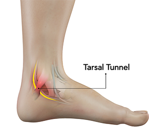
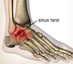
Sinus tarsi syndrome is a condition that affects the foot and ankle. It is characterized by pain and discomfort in the sinus tarsi, which is a small bony canal located between the talus bone (ankle bone) and the calcaneus bone (heel bone). The sinus tarsi serves as a passage for nerves, blood vessels, and ligaments that are important for foot and ankle function.

Sinus tarsi syndrome is typically caused by injury or trauma to the foot and ankle, such as ankle sprains, repetitive overuse, or chronic instability. Other potential causes can include arthritis, ligamentous laxity, or anatomical variations that lead to compression or irritation of the nerves or tissues within the sinus tarsi.

Symptoms of sinus tarsi syndrome may include:

  1. Pain: Pain in the lateral (outer) aspect of the foot, specifically in the area of the sinus tarsi, is the hallmark symptom of this condition. The pain may be sharp or dull and may worsen with weight-bearing activities or prolonged standing.
  2. Swelling: Swelling around the sinus tarsi may be present, although it is usually mild compared to other foot and ankle conditions.
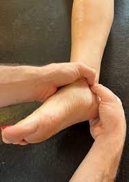
  3. Tenderness: Tenderness to touch over the sinus tarsi area may be present, and pressure on the area may exacerbate the pain.
  4. Instability: Some patients with sinus tarsi syndrome may experience a feeling of instability or a sense of “giving way” in the foot or ankle.
  5. Limited range of motion: Reduced range of motion in the ankle joint may be observed, particularly with movements that involve inversion (inward rolling) or eversion (outward rolling) of the foot.

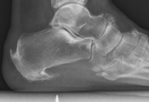
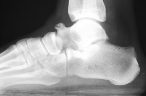
Diagnosis of sinus tarsi syndrome typically involves a thorough clinical evaluation by a healthcare provider, including a physical examination, assessment of medical history, and imaging studies such as X-rays or MRI to rule out other potential causes of foot and ankle pain.

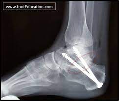
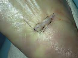
Treatment for sinus tarsi syndrome may include conservative measures such as rest, ice, compression, and elevation (RICE), nonsteroidal anti-inflammatory drugs (NSAIDs), orthotics or shoe modifications, physical therapy, and activity modification. In some cases, corticosteroid injections may be used to reduce inflammation and alleviate pain. If conservative measures are not effective, more advanced treatments such as extracorporeal shockwave therapy, prolotherapy, or platelet-rich plasma (PRP) injections may be considered. In rare cases, surgical intervention may be necessary to address any underlying structural issues or persistent symptoms.

It is important to consult with a qualified healthcare provider for an accurate diagnosis and appropriate treatment plan if you suspect you may have sinus tarsi syndrome or are experiencing foot and ankle pain.

Forestier’s disease

Forestier’s disease can affect various parts of the body, including the spine, pelvis, hip, and knee, but it is less common for it to affect the foot. However, in rare cases, Forestier’s disease can cause abnormal bone growth in the foot, leading to stiffness, pain, and difficulty with movement.

The symptoms of Forestier’s disease in the foot may include:

  • Stiffness and limited range of motion in the affected joint(s)
  • Pain that worsens with activity
  • Swelling or tenderness around the affected area
  • Deformity or abnormal bone growth in the foot

Treatment for Forestier’s disease in the foot may include nonsteroidal anti-inflammatory drugs (NSAIDs) for pain and inflammation, physical therapy to improve range of motion and mobility, and in severe cases, surgery to remove or reshape the abnormal bone growth. It’s important to consult with a healthcare provider for proper diagnosis and treatment of Forestier’s disease in the foot.

Subtalar joint arthrodesis

A subtalar joint arthrodesis is a surgical procedure in which the subtalar joint, which is located between the heel bone (calcaneus) and the ankle bone (talus), is fused together to create a single, solid bone. This procedure is typically performed to treat conditions such as severe arthritis, instability, or deformity of the subtalar joint.

During the procedure, the surgeon makes an incision on the side of the foot and removes the damaged cartilage from the subtalar joint. The bones are then positioned in the desired alignment and held in place with screws or other hardware. Over time, the bones grow together and fuse into a single, solid bone.

Subtalar joint arthrodesis is typically performed under general anesthesia and requires a period of immobilization in a cast or brace. Physical therapy is also recommended to help regain strength and mobility in the affected foot. While this procedure can be highly effective in treating certain conditions, it does limit the range of motion in the foot and ankle and may lead to an increased risk of arthritis in adjacent joints over time. It is important to discuss the potential risks and benefits of subtalar joint arthrodesis with a qualified healthcare professional before undergoing the procedure.

Instep fasciotomy (plantar fasciitis surgery)

An instep plantar fasciotomy is a surgical procedure that involves cutting a portion of the plantar fascia ligament in the foot to relieve tension and pain. The plantar fascia is a thick band of tissue that runs along the bottom of the foot, connecting the heel bone to the toes. When this tissue becomes inflamed or irritated, it can cause a condition called plantar fasciitis, which is characterized by pain and stiffness in the heel and sole of the foot.

During an instep plantar fasciotomy, the surgeon makes a small incision on the top of the foot, near the base of the toes, and cuts a portion of the plantar fascia ligament. This reduces tension on the ligament and relieves pressure on the affected area. The incision is typically closed with stitches, and the foot is immobilized in a cast or boot for several weeks to allow for proper healing.

While instep plantar fasciotomy can be an effective treatment for plantar fasciitis, it is typically reserved for cases that have not responded to non-surgical treatments, such as rest, stretching, and physical therapy. As with any surgical procedure, there are risks and potential complications associated with instep plantar fasciotomy, and it is important to discuss these with a qualified healthcare professional before undergoing the procedure.

Valleix’s sign

Valleix’s sign is another diagnostic test that can be used to assess nerve function in the peripheral nervous system. It involves applying pressure or pinching along the course of a nerve to identify areas of tenderness or pain.

In the case of tarsal tunnel syndrome, the Valleix sign can be used to identify areas of nerve irritation or damage along the course of the posterior tibial nerve. The healthcare provider will apply pressure or pinch along the nerve pathway, starting at the ankle and moving up the leg, while asking the patient if they feel any pain or discomfort.

If the patient experiences pain or tenderness along the course of the posterior tibial nerve, this can be an indication of nerve irritation or damage. However, like Tinel’s sign, the Valleix sign is not always definitive, and additional diagnostic tests may be needed to confirm the diagnosis.

In general, a combination of diagnostic tests and a thorough medical history and physical exam are used to diagnose tarsal tunnel syndrome. Treatment may involve rest, ice, physical therapy, and other conservative measures, and in some cases, surgery may be necessary to relieve pressure on the affected nerve.

Progressive collapsing flatfoot deformity – Classification system

Progressive collapsing flatfoot deformity can be classified according to the severity of the deformity, as well as the underlying cause. There are several classification systems used by healthcare providers to describe the different stages of the condition. Here are two common classification systems:

  1. Johnson and Strom’s classification system:
  • Stage I: The foot is flat when bearing weight, but the arch returns when the foot is lifted off the ground.
  • Stage II: The arch does not return when the foot is lifted off the ground, but the foot is still flexible.
  • Stage III: The foot is rigid, and there is significant arthritis and deformity.
  1. Myerson’s classification system:
  • Stage I: The hindfoot is in a valgus position (tilted outward) and the arch is slightly flattened.
  • Stage II: The hindfoot is in a valgus position, the arch is more severely flattened, and there is evidence of instability in the ankle joint.
  • Stage III: The hindfoot is in a valgus position, the arch is severely flattened, and there is significant ankle joint arthritis.

The classification system used will depend on the healthcare provider’s preference and the specific needs of the patient.

Why does my heel hurt?

Heel pain is a common complaint that can be caused by a number of different conditions. Here are some of the most common causes of heel pain:

  1. Plantar Fasciitis: This is a condition where the plantar fascia, a band of tissue that runs along the bottom of the foot, becomes inflamed. This can cause pain in the heel or arch of the foot, especially when taking the first steps in the morning or after periods of inactivity.
  2. Heel Spurs: A heel spur is a bony growth that forms on the heel bone, often as a result of long-term plantar fasciitis. The spur itself is not painful, but it can cause pain by irritating the surrounding tissues.
  3. Achilles Tendinitis: This is a condition where the Achilles tendon, which connects the calf muscles to the heel bone, becomes inflamed. This can cause pain and stiffness in the heel or back of the ankle.
  4. Bursitis: Bursitis is a condition where the bursae, small fluid-filled sacs that cushion the joints, become inflamed. This can cause pain in the heel or other parts of the foot.
  5. Stress Fracture: A stress fracture is a small crack in the bone that can be caused by overuse or repetitive impact. This can cause pain in the heel or other parts of the foot.
  6. Nerve Impingement: A nerve in the foot can become pinched or compressed, leading to pain in the heel or other parts of the foot.

If you are experiencing heel pain, it’s important to see a healthcare provider or a podiatrist to determine the underlying cause of the problem. They can help diagnose the condition and recommend appropriate treatments, such as stretching exercises, orthotics, or physical therapy, to help relieve the pain and improve your mobility.

Tibialis anterior tendinopathy

Tibialis anterior tendinopathy is a condition that affects the tibialis anterior tendon, which is located at the front of the ankle and helps to lift the foot upwards. This condition is caused by overuse or repetitive strain on the tendon, and can result in pain, swelling, and weakness in the affected area.

Symptoms of tibialis anterior tendinopathy include pain and tenderness along the front of the ankle, swelling and stiffness in the area, and difficulty lifting the foot upwards. The pain may be worse during physical activity or after prolonged periods of standing or walking.

Treatment for tibialis anterior tendinopathy typically involves rest, ice, compression, and elevation (RICE) to reduce pain and swelling. Non-steroidal anti-inflammatory drugs (NSAIDs) may also be recommended to relieve pain and inflammation. Physical therapy may be recommended to help strengthen the muscles and improve flexibility in the affected area. In severe cases, surgery may be required to repair or replace the damaged tendon.

Prevention of tibialis anterior tendinopathy involves wearing proper footwear and using orthotics or inserts to support the feet and reduce stress on the tendon. Gradually increasing activity levels and avoiding sudden changes in activity levels can also help prevent this condition. It is important to seek medical attention if you experience any symptoms of tibialis anterior tendinopathy, as early diagnosis and treatment can help prevent further damage and improve outcomes.

Tarsal tunnel syndrome

Tarsal tunnel syndrome is a condition in which the tibial nerve, which runs down the leg and into the foot, is compressed as it passes through a narrow tunnel in the ankle called the tarsal tunnel. This can result in pain, tingling, and numbness in the foot and ankle, as well as weakness and muscle atrophy in severe cases.

The symptoms of tarsal tunnel syndrome may be similar to those of other conditions affecting the foot and ankle, such as plantar fasciitis or nerve entrapment in the foot. Therefore, a thorough physical examination, along with imaging tests such as X-rays, MRI, or nerve conduction studies, may be necessary to confirm the diagnosis.

Conservative treatment options for tarsal tunnel syndrome may include rest, icing, physical therapy, and wearing supportive footwear or orthotics. Non-steroidal anti-inflammatory drugs (NSAIDs) may also be prescribed to help relieve pain and inflammation.

In more severe cases or when conservative treatments are not effective, surgery may be necessary to release the pressure on the tibial nerve by removing any structures that may be compressing it.

Early diagnosis and treatment are important for preventing long-term complications such as chronic pain, muscle atrophy, and nerve damage. If you experience persistent foot or ankle pain or notice any unusual symptoms, it is important to seek medical attention to determine the underlying cause and appropriate treatment.

Tarsal coalition

Tarsal coalition is a condition where two or more bones in the foot are abnormally fused together, resulting in limited mobility and pain. It is typically a congenital condition, meaning that it is present at birth, and it often becomes symptomatic during adolescence.

The most common type of tarsal coalition involves the calcaneus bone (heel bone) and the talus bone (bone between the heel bone and the ankle bone), but it can also occur between other bones in the foot.

Symptoms of tarsal coalition may include pain, stiffness, and limited range of motion in the affected foot, particularly during physical activity or standing for prolonged periods. In some cases, the affected foot may also appear to be flat or have a high arch.

Diagnosis of tarsal coalition may involve a physical exam, imaging tests such as X-rays or CT scans, and sometimes, bone scans or MRI.

Treatment for tarsal coalition may depend on the severity of the symptoms and the degree of fusion between the affected bones. Conservative treatment options may include rest, immobilisation with a cast or brace, physical therapy, and non-steroidal anti-inflammatory drugs (NSAIDs) to relieve pain and inflammation.

In more severe cases, surgery may be necessary to remove the abnormal bone or to fuse the affected bones together in a more functional position.

It is important to seek medical attention if you experience persistent foot pain or notice any unusual symptoms, as untreated tarsal coalition can lead to long-term complications such as chronic pain and arthritis.