Category: Surgical

Lapidus procedure

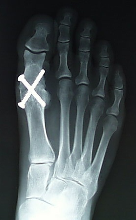
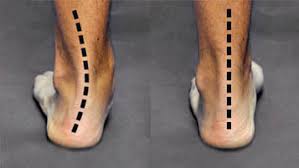
The Lapidus procedure is a surgical procedure used to treat hallux valgus, a condition in which the big toe drifts away from the midline of the foot, causing a bunion. It involves fusing the first metatarsal bone to the medial cuneiform bone in the midfoot to correct the alignment of the bones and reduce the deformity. This procedure is typically reserved for cases of severe hallux valgus or for patients who have not responded to more conservative treatments.

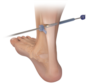
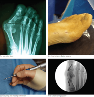
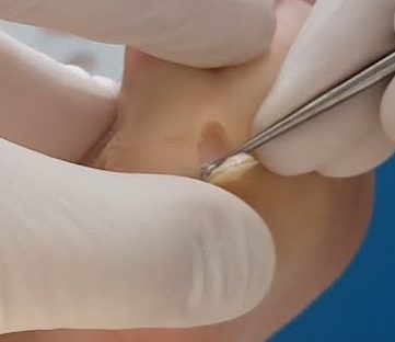
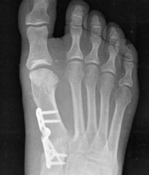
During the procedure, the surgeon makes an incision on the top of the foot and removes a small piece of bone from the base of the first metatarsal. The metatarsal bone is then repositioned and fixed in place with screws or a plate to hold it in the desired alignment. Over time, the bones grow together and form a solid fusion, which helps to stabilize the midfoot and reduce the severity of the bunion.

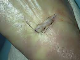
After the surgery, the patient may need to wear a cast or brace for several weeks to protect the foot and allow the bones to fuse together. Physical therapy may also be recommended to help improve strength, flexibility, and range of motion in the affected foot. While the Lapidus procedure can be highly effective in correcting hallux valgus, it does require a period of immobilization and recovery, and may have some potential risks and complications, such as non-union, nerve injury, or infection. It is important to discuss the potential risks and benefits of the Lapidus procedure with a qualified healthcare professional before undergoing the procedure.

First metatarsophalangeal joint arthrodesis

A first metatarsophalangeal joint (1st MTPJ) arthrodesis is a surgical procedure that involves fusing the bones of the big toe (metatarsal) and the first bone of the toe (phalanx) together to create a solid joint. This procedure is typically performed to treat advanced arthritis, instability, or deformity of the 1st MTPJ.

During the procedure, the surgeon makes an incision on the top of the foot and removes the damaged joint surfaces of the metatarsal and phalanx bones. The bones are then placed in the desired alignment and held in place with screws, pins, or plates until they fuse together. Over time, the bones grow together and form a solid joint, which can help relieve pain and improve the stability and function of the foot.

After the surgery, the foot is immobilized in a cast or boot for several weeks, and physical therapy may be recommended to help regain strength and mobility in the affected foot. While 1st MTPJ arthrodesis can be highly effective in treating certain conditions, it does limit the range of motion in the big toe and may affect the gait or balance of the patient. It is important to discuss the potential risks and benefits of 1st MTPJ arthrodesis with a qualified healthcare professional before undergoing the procedure.

Subtalar joint arthrodesis

A subtalar joint arthrodesis is a surgical procedure in which the subtalar joint, which is located between the heel bone (calcaneus) and the ankle bone (talus), is fused together to create a single, solid bone. This procedure is typically performed to treat conditions such as severe arthritis, instability, or deformity of the subtalar joint.

During the procedure, the surgeon makes an incision on the side of the foot and removes the damaged cartilage from the subtalar joint. The bones are then positioned in the desired alignment and held in place with screws or other hardware. Over time, the bones grow together and fuse into a single, solid bone.

Subtalar joint arthrodesis is typically performed under general anesthesia and requires a period of immobilization in a cast or brace. Physical therapy is also recommended to help regain strength and mobility in the affected foot. While this procedure can be highly effective in treating certain conditions, it does limit the range of motion in the foot and ankle and may lead to an increased risk of arthritis in adjacent joints over time. It is important to discuss the potential risks and benefits of subtalar joint arthrodesis with a qualified healthcare professional before undergoing the procedure.

Instep fasciotomy (plantar fasciitis surgery)

An instep plantar fasciotomy is a surgical procedure that involves cutting a portion of the plantar fascia ligament in the foot to relieve tension and pain. The plantar fascia is a thick band of tissue that runs along the bottom of the foot, connecting the heel bone to the toes. When this tissue becomes inflamed or irritated, it can cause a condition called plantar fasciitis, which is characterized by pain and stiffness in the heel and sole of the foot.

During an instep plantar fasciotomy, the surgeon makes a small incision on the top of the foot, near the base of the toes, and cuts a portion of the plantar fascia ligament. This reduces tension on the ligament and relieves pressure on the affected area. The incision is typically closed with stitches, and the foot is immobilized in a cast or boot for several weeks to allow for proper healing.

While instep plantar fasciotomy can be an effective treatment for plantar fasciitis, it is typically reserved for cases that have not responded to non-surgical treatments, such as rest, stretching, and physical therapy. As with any surgical procedure, there are risks and potential complications associated with instep plantar fasciotomy, and it is important to discuss these with a qualified healthcare professional before undergoing the procedure.

Gastrocnemius recession (calf release)

A gastrocnemius recession, also known as gastrocnemius lengthening or calf muscle release, is a surgical procedure in which the gastrocnemius muscle is partially cut to relieve tension and tightness in the calf. This procedure is typically performed to treat a condition called equinus deformity, which is characterized by limited ankle dorsiflexion (the ability to pull the foot upward toward the shin).

During the procedure, a small incision is made in the skin over the gastrocnemius muscle, and the muscle is partially cut or released from its attachment to the heel bone (calcaneus). This allows the muscle to stretch more easily and reduces tension on the Achilles tendon and plantar fascia.

Gastrocnemius recession is often performed in combination with other procedures, such as Achilles tendon lengthening or plantar fascia release, to address the underlying cause of equinus deformity. Recovery from the procedure typically involves a period of immobilization and physical therapy to regain strength and flexibility in the affected leg. As with any surgery, there are risks and potential complications associated with gastrocnemius recession, and it is important to discuss these with a qualified healthcare professional before undergoing the procedure.

Subtalar joint implants for flat feet

Subtalar joint implants may be recommended for patients with flat feet or other foot deformities that affect the subtalar joint. The subtalar joint is located in the ankle and connects the talus bone to the calcaneus bone. It plays an important role in foot movement and stability.

A subtalar joint implant is a small device that is surgically implanted into the subtalar joint to help stabilize and realign the bones of the foot. The implant may be made of metal, plastic, or other materials and is designed to mimic the natural movement of the subtalar joint.

Subtalar joint implants may be recommended for patients with flat feet who have not responded to conservative treatments such as orthotics, physical therapy, or medication. The implant can help reduce pain, improve foot function, and prevent further damage to the foot.

However, like any surgical procedure, subtalar joint implant surgery carries some risks, including infection, bleeding, and nerve damage. Patients may also experience some pain and swelling after the surgery, and may need to wear a cast or brace for several weeks while the foot heals.

If you are considering subtalar joint implant surgery for flat feet, it is important to discuss the risks and benefits of the procedure with your healthcare provider to determine if it is the right option for you.

Preventing infection in foot and ankle surgery

Preventing infection in foot and ankle surgery is crucial to ensure proper healing and recovery. Here are some ways to prevent infection in foot and ankle surgery:

  1. Preoperative antibiotics: Administering antibiotics before surgery can help reduce the risk of infection. The type and duration of antibiotics may vary depending on the patient’s health status and the type of surgery.
  2. Sterilisation: All surgical instruments, equipment, and materials must be sterilised before use to eliminate any potential sources of infection.
  3. Proper surgical technique: Following proper surgical technique, including maintaining a sterile field, minimising tissue trauma, and using appropriate wound closure techniques, can help prevent infection.
  4. Skin preparation: Preoperative skin preparation, such as using antiseptic solutions, can help reduce the number of bacteria on the skin, which can decrease the risk of infection.
  5. Patient education: Educating the patient about proper wound care and postoperative hygiene can help reduce the risk of infection. Patients should be advised to keep the wound clean and dry and to report any signs of infection, such as redness, swelling, or drainage, to their healthcare provider immediately.
  6. Proper ventilation: The operating room should be well-ventilated to help reduce the risk of airborne contamination.
  7. Immunisation: Patients should be immunised against relevant pathogens before surgery to help reduce the risk of infection.

By following these steps, the risk of infection can be significantly reduced, leading to better outcomes for patients undergoing foot and ankle surgery.

Minimally-invasive (keyhole) foot surgery

Minimally-invasive (keyhole) foot surgery is a type of foot surgery that is performed using small incisions and specialised instruments. It is also known as percutaneous surgery or minimally invasive surgery (MIS). This type of surgery is designed to minimise tissue trauma, reduce postoperative pain and swelling, and speed up recovery time compared to traditional open surgery.

During the surgery, the surgeon will make small incisions in the skin and insert specialised instruments, such as cameras and small cutting tools, to perform the procedure. Because the incisions are smaller, there is less tissue trauma and bleeding, and patients typically experience less pain and scarring.

Minimally-invasive foot surgery can be used to treat a variety of foot and ankle conditions, including bunions, hammertoes, plantar fasciitis, neuromas, and Achilles tendon injuries. The suitability of this type of surgery depends on the individual patient’s condition and medical history, as well as the surgeon’s experience and expertise.

Benefits of minimally-invasive foot surgery include a shorter recovery time, reduced pain and swelling, less scarring, and a lower risk of infection. However, there are also potential risks and complications associated with any type of surgery, so it is important to discuss the benefits and risks with your doctor before making a decision about treatment.

Curettage for warts

Wart curettage is a medical procedure used to remove warts by scraping or cutting them away from the skin. The procedure is typically performed using a small, sharp instrument called a curette. The area is first numbed with a local anaesthetic, and then the wart is scraped or cut away using the curette.

After the procedure, the wound is typically covered with a bandage or dressing. Pain relief medication may be prescribed, and patients are usually advised to avoid getting the area wet for a few days after the procedure.

Wart curettage is generally considered a safe and effective treatment for warts, but there is a risk of scarring or infection, particularly if the wound is not properly cared for after the procedure. It is important to follow your doctor’s instructions carefully and to contact them if you experience any signs of infection, such as redness, swelling, or fever.

Cosmetic foot surgery

Cosmetic foot surgery, also known as aesthetic foot surgery, is a type of elective surgery that is performed for purely cosmetic reasons rather than medical reasons. The goal of cosmetic foot surgery is to improve the appearance of the foot or correct a perceived imperfection, such as bunions, hammertoes, or other cosmetic issues.

Some of the most common types of cosmetic foot surgery include bunion surgery, hammertoe surgery, toe shortening or lengthening, and fat pad augmentation. These procedures are typically performed on an outpatient basis and may involve local anaesthesia, sedation, or general anaesthesia, depending on the specific procedure and the patient’s needs.

While cosmetic foot surgery can improve the appearance of the feet, it is important to remember that any surgery involves risks, including infection, bleeding, nerve damage, and poor healing. It is important to discuss the potential risks and benefits of any cosmetic foot surgery with a qualified and experienced surgeon before deciding to undergo the procedure.