Category: Cancer

Synovial sarcoma

Synovial sarcoma is a rare type of soft tissue cancer that typically arises in the arms or legs, but can also occur in other parts of the body such as the head and neck, trunk, or abdomen. Despite its name, synovial sarcoma does not originate in the synovial tissue of joints, but rather from cells that resemble synovial cells.

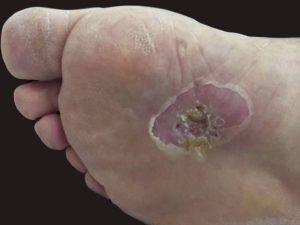
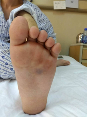
The cause of synovial sarcoma is not well understood, but genetic mutations and environmental factors may play a role. Symptoms of synovial sarcoma may include a painless lump or swelling, stiffness, and difficulty moving the affected area.

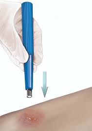
Diagnosis of synovial sarcoma may involve a physical exam, imaging tests such as X-rays or MRI, and a biopsy to confirm the presence of cancerous cells. Treatment for synovial sarcoma usually involves surgery to remove the tumour, followed by radiation therapy and chemotherapy. In some cases, amputation may be necessary if the tumour is too large or has spread extensively.

The prognosis for synovial sarcoma varies depending on the size and location of the tumour, as well as the stage of the cancer at the time of diagnosis. Early detection and treatment can improve the chances of successful treatment and long-term survival. It is important to seek medical attention if you notice any unusual lumps or growths on your body, particularly if they are painful or persist for more than a few weeks.

Squamous cell carcinoma

Squamous cell carcinoma (SCC) is a type of skin cancer that develops in the squamous cells that make up the outer layer of the skin. SCC can occur anywhere on the body, but it is most commonly found on areas of the skin that have been exposed to the sun, such as the face, neck, hands, and arms. Here are some key facts about squamous cell carcinoma:

Causes:

  • Exposure to ultraviolet (UV) radiation from the sun or tanning beds is the primary cause of squamous cell carcinoma.
  • Other risk factors include fair skin, a history of frequent sunburns, a weakened immune system, exposure to certain chemicals or toxins, and a history of previous skin cancer.

Symptoms:

  • A firm, red bump or flat, scaly patch on the skin that may be crusty or bleed easily.
  • An open sore that does not heal or heals and then returns.
  • A growth that looks like a wart or raised, rough patch on the skin.

Diagnosis:

  • A biopsy is the only way to definitively diagnose squamous cell carcinoma.
  • During a biopsy, a small sample of the affected tissue is removed and examined under a microscope to determine if cancerous cells are present.

Treatment:

  • Treatment for squamous cell carcinoma typically involves surgical removal of the cancerous tissue.
  • Mohs surgery, which involves removing thin layers of tissue and examining them under a microscope until all the cancerous cells are gone, is a common surgical technique for treating squamous cell carcinoma.
  • Other treatment options may include radiation therapy, topical chemotherapy, or immunotherapy.

Prognosis:

  • Squamous cell carcinoma is usually curable when detected and treated early.
  • If left untreated, squamous cell carcinoma can grow deeper into the skin and spread to other parts of the body, potentially becoming life-threatening.

Skin cancer of the foot

Skin cancer can occur on any part of the body, including the foot and ankle. The most common types of skin cancer that affect the feet are basal cell carcinoma, squamous cell carcinoma, and melanoma.

Basal cell carcinoma and squamous cell carcinoma are the most common types of skin cancer and are usually caused by sun exposure. They typically appear as a small, raised, or flat bump that is pink, red, or white in colour. They may also have a scaly or crusted surface.

Melanoma is a less common but more serious type of skin cancer that can spread quickly to other parts of the body. It typically appears as a dark, irregularly shaped spot or mole that may bleed or itch.

To prevent skin cancer on the foot and ankle, it is important to wear proper footwear that provides adequate sun protection, such as shoes with a closed toe and a thick sole. Additionally, use sunscreen on exposed areas of the foot and ankle, and avoid prolonged sun exposure.

If you notice any unusual changes in the skin on your feet or ankles, such as a new growth, change in colour or texture, or persistent sore or ulcer, it is important to see a healthcare professional for evaluation. Treatment for skin cancer may include surgical removal of the affected area, radiation therapy, or chemotherapy, depending on the type and severity of the cancer. Early detection and treatment are important for the best possible outcome.

Malignant melanoma of the foot

Malignant melanoma is a type of skin cancer that can develop on the foot. It can occur on any part of the foot, including the soles, nails, and between the toes. The signs and symptoms of melanoma of the foot include:

  1. A new mole, bump, or spot on the foot that is changing in size, shape, or colour
  2. A sore that does not heal
  3. A pigmented band that runs under the nail and extends onto the adjacent skin
  4. A dark spot or streak that grows in size or changes in colour
  5. A nodule, lump, or bump on the foot that is not painful but keeps growing

If you notice any of these signs or symptoms on your foot, you should see a podiatrist or dermatologist for evaluation. Early detection and treatment are important for the successful management of melanoma of the foot.

Kaposi’s sarcoma

Kaposi’s sarcoma (KS) is a rare type of cancer that affects the cells that line the blood vessels or lymphatic vessels. KS can occur in several forms, including classic, endemic, iatrogenic, and epidemic (or AIDS-related) KS. Here are some key facts about Kaposi’s sarcoma:

Causes:

  • Kaposi’s sarcoma is caused by a type of herpes virus called human herpesvirus 8 (HHV-8), also known as Kaposi’s sarcoma-associated herpesvirus (KSHV).
  • Risk factors for developing Kaposi’s sarcoma include having a weakened immune system, being infected with HIV, and being of Mediterranean or African descent.

Symptoms:

  • Kaposi’s sarcoma usually appears as patches or nodules on the skin, which may be red, purple, or brown.
  • The lesions may be flat or raised and may itch or be painful.
  • KS can also affect other parts of the body, such as the lungs, gastrointestinal tract, and lymph nodes.

Diagnosis:

  • A biopsy is the only way to definitively diagnose Kaposi’s sarcoma.
  • During a biopsy, a small sample of the affected tissue is removed and examined under a microscope to determine if cancerous cells are present.

Treatment:

  • Treatment for Kaposi’s sarcoma depends on the type and extent of the cancer, as well as the patient’s overall health.
  • Treatments may include radiation therapy, chemotherapy, or targeted therapy.
  • Antiretroviral therapy (ART) may also be used to treat Kaposi’s sarcoma in people who are infected with HIV.

Prognosis:

  • The prognosis for Kaposi’s sarcoma varies depending on the type and extent of the cancer and the patient’s overall health.
  • In people with AIDS-related KS, the prognosis is generally worse than in other forms of the disease, as the cancer may be more aggressive and difficult to treat. However, with appropriate treatment, many people with Kaposi’s sarcoma are able to manage the disease and live for many years.

Chemotherapy-induced foot problems

Chemotherapy can cause various side effects on the feet, including:

  1. Peripheral neuropathy: Chemotherapy can cause damage to the peripheral nerves that control the sensations and movements of the feet. This can result in symptoms such as numbness, tingling, burning, and pain in the feet, which can affect mobility and balance.
  2. Weakness and fatigue: Chemotherapy can also cause weakness and fatigue in the muscles of the feet, making it difficult to stand, walk, or perform other physical activities.
  3. Infection: Chemotherapy can weaken the immune system, making it more susceptible to infections, including foot infections.
  4. Nail changes: Chemotherapy can cause changes in the appearance and texture of the nails, such as discolouration, ridges, and brittleness.
  5. Swelling: Chemotherapy can cause swelling in the feet and ankles, which can be uncomfortable and interfere with mobility.

It is important for cancer patients undergoing chemotherapy to maintain good foot hygiene, wear comfortable shoes that fit properly, and report any changes in foot health to their healthcare provider. They may also benefit from physical therapy, orthotics, and other supportive measures to manage foot symptoms and maintain mobility.

Biopsy

A biopsy is a medical procedure in which a small sample of tissue is taken from a suspicious or abnormal area on the foot for further examination under a microscope. This procedure can help diagnose the cause of the lesion, such as infection, inflammation, or cancer.

The biopsy can be performed using various methods, depending on the size and location of the lesion. Some common techniques include:

  1. Punch biopsy: A small, circular instrument is used to remove a cylindrical sample of skin and tissue from the lesion.
  2. Incisional biopsy: A surgical knife is used to remove a small wedge-shaped section of the lesion.
  3. Excisional biopsy: The entire lesion is removed, along with some surrounding healthy tissue.
  4. Needle biopsy: A thin needle is inserted into the lesion to remove a small sample of cells.

The type of biopsy performed will depend on the location and size of the lesion, as well as the suspected diagnosis. In some cases, the biopsy may be performed under local anaesthesia to minimise discomfort.

After the biopsy, the sample is sent to a laboratory for analysis. The results of the biopsy can help guide further treatment and management of the foot lesion. Your doctor will discuss the results of the biopsy with you and recommend appropriate next steps based on the diagnosis.

Basal cell carcinoma (BCC)

Basal cell carcinoma (BCC) is a common type of skin cancer that arises from the basal cells in the outermost layer of the skin. BCC can occur anywhere on the body but is most often found on areas that have been exposed to the sun, such as the face, neck, and hands. Here are some key facts about basal cell carcinoma:

Causes:

  • Exposure to ultraviolet (UV) radiation from the sun or tanning beds is the primary cause of basal cell carcinoma.
  • Other risk factors include fair skin, a history of frequent sunburns, a weakened immune system, and a history of previous skin cancer.

Symptoms:

  • A pearly or waxy bump on the skin that may be flesh-coloured, pink, or brown.
  • A flat, scaly, or crusty patch on the skin that may be red or brown.
  • A sore that does not heal or heals and then returns.

Diagnosis:

  • A biopsy is the only way to definitively diagnose basal cell carcinoma.
  • During a biopsy, a small sample of the affected tissue is removed and examined under a microscope to determine if cancerous cells are present.

Treatment:

  • Treatment for basal cell carcinoma typically involves surgical removal of the cancerous tissue.
  • Mohs surgery, which involves removing thin layers of tissue and examining them under a microscope until all the cancerous cells are gone, is a common surgical technique for treating basal cell carcinoma.
  • Other treatment options may include radiation therapy, topical chemotherapy, or immunotherapy.

Prognosis:

  • Basal cell carcinoma is usually curable when detected and treated early.
  • If left untreated, basal cell carcinoma can grow deeper into the skin and spread to other parts of the body, potentially becoming life-threatening. However, this is rare.