Category: Paediatrics

Clinodactyly

Clinodactyly is a medical term describing the curvature of a digit (a finger or toe) in the plane of the palm, most commonly the fifth finger (the “little finger”) towards the adjacent fourth finger (the “ring finger”).

It is a fairly common isolated anomaly which often goes unnoticed, but also occurs in combination with other abnormalities in certain genetic syndromes.[1] The term is from the Ancient Greek κλίνειν klínein ‘to bend’ and δάκτυλος dáktulos ‘digit’.

Skew foot

Skewfoot, also known as oblique foot or serpentine foot, is a rare foot deformity that can be present at birth or develop over time. It is characterized by an abnormal position of the heel bone (calcaneus) and the midfoot bones, resulting in a foot that is twisted and tilted inwards.

The exact cause of skewfoot is not fully understood, but it is believed to be related to abnormal development of the bones and joints in the foot. The condition can also be associated with other foot and leg problems, such as flat feet, high arches, and hip dysplasia.

The symptoms of skewfoot can include pain, stiffness, and difficulty walking or standing for long periods of time. Treatment options may include wearing orthotics or special shoes, physical therapy to improve foot alignment and range of motion, or surgery to correct the deformity in severe cases.

If you suspect you have skewfoot, it is recommended that you consult with a podiatrist or orthopedic surgeon who specializes in foot and ankle conditions for a proper diagnosis and treatment plan.

Polydactyly

Polydactyly is a genetic condition characterized by the presence of extra fingers or toes on a person’s hands or feet. Instead of the typical five digits on each hand or foot, a person with polydactyly may have six, seven, or even more digits. The extra digits can be fully formed and functional or they may be small, non-functional nubs.

Polydactyly can be inherited in an autosomal dominant pattern, meaning that a child has a 50% chance of inheriting the condition if one parent carries the gene. In some cases, however, polydactyly can occur spontaneously as a random mutation.

Polydactyly is usually not a serious medical condition, and many people with the condition are able to live normal, healthy lives without any problems. In some cases, however, surgery may be necessary to remove the extra digits if they are causing problems with mobility or other issues.

Tillaux fracture

A Tillaux fracture is a specific type of ankle fracture that occurs in children and adolescents. It is caused by a twisting injury to the ankle and is characterized by a fracture of the lateral aspect of the tibial epiphysis.

The tibial epiphysis is the area of developing bone at the end of the tibia that contributes to the growth of the bone. The lateral aspect of the tibial epiphysis is where the fibula bone attaches to the tibia. When a twisting force is applied to the ankle, it can cause the fibula to pull on the lateral aspect of the tibial epiphysis, resulting in a Tillaux fracture.

Symptoms of a Tillaux fracture may include pain, swelling, and difficulty bearing weight on the affected foot. Treatment typically involves immobilization of the ankle with a cast or brace to allow the fracture to heal. In some cases, surgery may be necessary to realign and stabilize the fractured bone.

If left untreated or improperly treated, a Tillaux fracture can lead to long-term complications such as chronic pain, instability of the ankle joint, and an increased risk of developing arthritis in the ankle. Early and appropriate treatment is important for the best possible outcome.

Köhler disease

Köhler disease, also known as osteochondrosis of the navicular bone, is a rare condition that affects the navicular bone in the foot. It is most commonly seen in children between the ages of 5 and 10 years old, and is more common in boys than girls.

The exact cause of Köhler disease is unknown, but it is believed to be related to a disruption in blood flow to the navicular bone. This can lead to a decrease in bone density and the development of small fractures in the bone.

Symptoms of Köhler disease typically include pain and swelling in the midfoot, particularly on the top of the foot. The affected foot may also appear flattened or widened, and there may be a limp or difficulty walking.

Treatment for Köhler disease usually involves rest and immobilization of the foot with a cast or brace to allow the bone to heal. Pain relievers may also be prescribed to manage discomfort. In rare cases, surgery may be necessary to remove damaged tissue or realign the bones in the foot. With appropriate treatment, most children with Köhler disease recover fully and have no long-term complications.

Salter-Harris classification for growth plate fractures

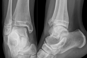
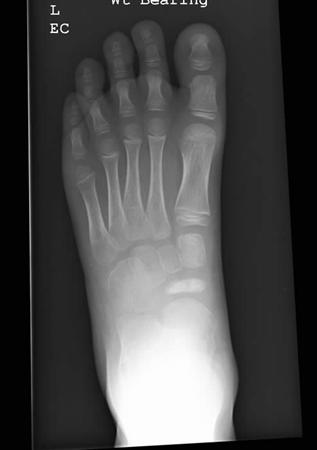
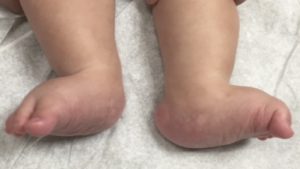
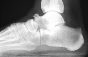
The Salter-Harris classification is a system used to classify fractures that involve the growth plate, also known as the epiphyseal plate, in pediatric patients. The growth plate is a cartilage-rich area at the ends of long bones that allows for bone growth and development.

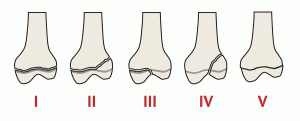
The Salter-Harris classification divides growth plate fractures into five categories, based on the location and extent of the fracture:

Type I: This is a transverse fracture that runs through the growth plate, separating the epiphysis (the end of the bone) from the metaphysis (the shaft of the bone). This is the most common type of growth plate fracture and is usually treated with immobilization and close monitoring.

Type II: This is an oblique fracture that runs through the growth plate and into the metaphysis. This type of fracture is also treated with immobilization and monitoring, and may require more frequent follow-up to ensure proper healing.

Type III: This is a fracture that runs through the growth plate and into the epiphysis. This type of fracture may require more aggressive treatment, such as surgery, to prevent long-term complications such as growth disturbances or joint deformities.

Type IV: This is a fracture that runs through the growth plate, the epiphysis, and the metaphysis. This type of fracture is relatively rare and may require surgical intervention to prevent long-term complications.

Type V: This is a crush injury to the growth plate that results in damage to the cells responsible for bone growth. This type of fracture is also relatively rare and may require surgical intervention to prevent growth disturbances.

The Salter-Harris classification is a useful tool for healthcare professionals in assessing and managing growth plate fractures in pediatric patients. Treatment options for growth plate fractures may include immobilization, closed reduction (manipulation of the bones to restore proper alignment), and surgery in some cases.

Overall, prompt and appropriate treatment of growth plate fractures is important to minimize the risk of long-term complications and ensure proper bone growth and development.

Vertical talus

Vertical talus, also known as congenital vertical talus or convex pes valgus, is a rare foot deformity in which the talus bone (the bone that connects the foot to the ankle) is abnormally positioned, causing the foot to be in a fixed and rigid upward position. Here are some key facts about vertical talus:

Causes:

  • The exact cause of vertical talus is not known, but it is believed to be a congenital condition that occurs during foetal development.
  • It may be associated with other genetic syndromes or abnormalities, such as arthrogryposis or spina bifida.

Symptoms:

  • Symptoms of vertical talus include a visible upward curve of the foot, with the heel tilted downward and the front of the foot pointing upward.
  • The foot may also be stiff and rigid, making it difficult or impossible to flex the ankle.
  • In some cases, there may be pain or discomfort in the foot or ankle.

Diagnosis:

  • A diagnosis of vertical talus is usually made based on a physical examination of the foot and the patient’s medical history.
  • X-rays or other imaging tests may be ordered to confirm the diagnosis and assess the severity of the deformity.

Treatment:

  • Treatment for vertical talus usually involves surgery to correct the position of the talus bone and realign the foot.
  • Depending on the severity of the deformity, surgery may involve releasing or lengthening tight or shortened tendons and ligaments, and/or placing pins or screws to hold the bones in the correct position.
  • In some cases, casting or bracing may be used before or after surgery to support the foot and aid in healing.

Prognosis:

  • With appropriate treatment, most cases of vertical talus can be corrected.
  • However, the success of treatment depends on the severity of the deformity and the age of the patient at the time of treatment.
  • Patients with vertical talus may need long-term monitoring and follow-up to ensure that the foot remains properly aligned and to address any future complications.

Toe walking

Toe walking is a condition where a person habitually walks on their toes or the balls of their feet, rather than with their heels on the ground. It is relatively common in infants and young children, but can also occur in older children and adults.

There are many possible causes of toe walking, including neurological conditions such as cerebral palsy, muscular dystrophy, and autism spectrum disorder. Some people may also toe walk due to tightness or shortening of the calf muscles, or as a result of a foot or ankle deformity.

In some cases, toe walking may not cause any problems and may even resolve on its own over time. However, if toe walking persists beyond the age of 2 or 3, or is accompanied by other symptoms such as pain, stiffness, or difficulty with balance or coordination, it is important to seek medical advice.

Treatment for toe walking may include physical therapy, stretching exercises, and wearing braces or special shoes to help correct the gait abnormality. In some cases, surgery may be recommended to lengthen or release tight muscles or tendons. The appropriate treatment will depend on the underlying cause of the toe walking and the individual’s age, health, and lifestyle factors.

How to spot children’s foot problems

As children grow and develop, it is important to pay attention to their foot health to ensure proper growth and function. Here are some tips for parents on how to spot kids’ foot problems:

  1. Pay attention to how your child walks and runs. Look for any limping, toe-walking, or other unusual gait patterns.
  2. Check your child’s shoes regularly to make sure they are not too tight or too loose, and that they are wearing shoes that fit well and provide proper support.
  3. Look for any changes in your child’s foot shape or arches, as these may be signs of developing foot problems.
  4. Pay attention to your child’s complaints of foot pain or discomfort, especially after physical activity or prolonged periods of standing.
  5. Check for any signs of skin irritation, blisters, or calluses on your child’s feet.
  6. If your child has flat feet, is overweight, or participates in high-impact sports, they may be at greater risk for developing foot problems. Be sure to monitor their foot health closely.
  7. Consider taking your child to see a podiatrist if you notice any persistent foot problems or concerns.

By paying attention to your child’s foot health and seeking appropriate care when necessary, you can help ensure they have healthy and happy feet as they grow and develop.

Tarsal coalition

Tarsal coalition is a condition where two or more bones in the foot are abnormally fused together, resulting in limited mobility and pain. It is typically a congenital condition, meaning that it is present at birth, and it often becomes symptomatic during adolescence.

The most common type of tarsal coalition involves the calcaneus bone (heel bone) and the talus bone (bone between the heel bone and the ankle bone), but it can also occur between other bones in the foot.

Symptoms of tarsal coalition may include pain, stiffness, and limited range of motion in the affected foot, particularly during physical activity or standing for prolonged periods. In some cases, the affected foot may also appear to be flat or have a high arch.

Diagnosis of tarsal coalition may involve a physical exam, imaging tests such as X-rays or CT scans, and sometimes, bone scans or MRI.

Treatment for tarsal coalition may depend on the severity of the symptoms and the degree of fusion between the affected bones. Conservative treatment options may include rest, immobilisation with a cast or brace, physical therapy, and non-steroidal anti-inflammatory drugs (NSAIDs) to relieve pain and inflammation.

In more severe cases, surgery may be necessary to remove the abnormal bone or to fuse the affected bones together in a more functional position.

It is important to seek medical attention if you experience persistent foot pain or notice any unusual symptoms, as untreated tarsal coalition can lead to long-term complications such as chronic pain and arthritis.