Category: E

Extra bones in the foot

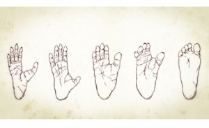
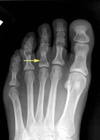
Extra bones in the foot are relatively common and can be present from birth or develop later in life. Some of the most common types of extra bones in the foot include:

  1. Accessory navicular: This is an extra bone that can be found on the inner side of the foot, near the ankle. It is present in about 10% of the population and can sometimes cause pain or discomfort.
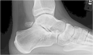
  2. Os trigonum: This is an extra bone that can be found behind the ankle bone (talus). It is present in about 10-15% of the population and may cause pain or limited mobility in some people.
  3. Hallux sesamoid: These are small bones that are embedded within the tendons of the big toe. They can sometimes become irritated or inflamed, causing pain or discomfort.
  4. Accessory cuneiform: These are extra bones that can be found in the middle of the foot, near the arch. They are relatively uncommon but can sometimes cause pain or deformity.
  5. Duplicated bones: In some cases, an extra bone may develop as a duplicate of an existing bone in the foot. This can cause problems if the duplicate bone causes pressure or impinges on nearby structures.

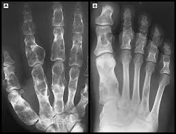
Extra bones in the foot are usually diagnosed through X-rays or other imaging tests. Treatment depends on the type and location of the extra bone, as well as the severity of symptoms. In some cases, non-surgical treatments such as rest, ice, and physical therapy may be recommended. In more severe cases, surgery may be necessary to remove the extra bone or address any associated problems. It is important to consult with a healthcare provider if you are experiencing foot pain or have concerns about extra bones in your foot.

How did the human foot evolve?

The human foot has evolved over millions of years, adapting to the demands of upright walking and running. Here are some key milestones in the evolution of the human foot:

  1. The earliest known hominids, such as Ardipithecus ramidus and Australopithecus afarensis, had feet that were adapted for climbing trees, with flexible toes and a grasping big toe that could grip branches.
  2. As early hominids began to walk upright on two legs, their feet gradually evolved to support this new mode of locomotion. The arches of the foot developed to absorb shock and provide stability, while the toes became shorter and less flexible.
  3. By the time of Homo erectus, around 2 million years ago, the foot had become more modern in appearance, with a longitudinal arch and a non-grasping big toe that was aligned with the other toes. This allowed for more efficient walking and running.
  4. The evolution of the foot continued with the appearance of Homo sapiens around 300,000 years ago. The arches of the foot became more pronounced, providing greater shock absorption and helping to distribute weight more evenly across the foot.
  5. In the last few thousand years, the human foot has undergone further changes due to cultural factors such as footwear. Shoes have altered the shape of the foot, leading to a higher incidence of foot problems such as bunions, hammertoes, and flat feet.

Overall, the human foot has evolved to support upright walking and running, with adaptations such as the arches of the foot and the alignment of the toes helping to distribute weight and absorb shock. However, cultural factors such as footwear can also impact the health and function of the foot.

Equinus

Equinus foot is a condition in which the ankle joint is limited in its ability to bend upward, also known as dorsiflexion. This condition can be caused by a variety of factors, including tight calf muscles, ankle joint stiffness or injury, or neurological conditions that affect the nerves and muscles of the foot and ankle.

Equinus foot can cause a number of symptoms, including difficulty walking or running, pain in the foot or ankle, and an increased risk of falls or injuries. In severe cases, equinus foot can lead to foot deformities or chronic ankle instability.

Treatment for equinus foot typically involves a combination of stretching exercises, physical therapy, and orthotic devices, such as heel lifts or ankle-foot orthoses (AFOs), that help to improve the ankle’s range of motion and support the foot and ankle during movement. In some cases, surgery may be necessary to correct the underlying structural problems that are causing equinus foot.

It is important to consult with a healthcare provider if you are experiencing symptoms of equinus foot or have difficulty moving your ankle joint. A healthcare provider can help to diagnose the underlying cause of the condition and develop an appropriate course of treatment based on your individual needs and medical history.

Erythrasma

Erythrasma is a bacterial skin infection caused by the bacterium Corynebacterium minutissimum. It is typically characterized by red, brown, or pinkish patches of skin that may be itchy or scaly. Here are some key facts about erythrasma:

Causes:

  • Erythrasma is caused by the bacterium Corynebacterium minutissimum.
  • The bacteria thrive in warm, moist environments, and the infection is most commonly found in the folds of the skin, such as the groin, armpits, and between the toes.

Symptoms:

  • Symptoms of erythrasma include red, brown, or pinkish patches of skin that may be itchy or scaly.
  • The patches may have a well-defined border and may be slightly raised or bumpy.
  • The infection may also cause a mild, musty odour.

Diagnosis:

  • A diagnosis of erythrasma is usually made based on the appearance of the skin lesions and the patient’s medical history.
  • A Wood’s lamp, which emits ultraviolet light, can be used to confirm the diagnosis by causing the affected skin to fluoresce.

Treatment:

  • Treatment for erythrasma typically involves topical or oral antibiotics to kill the bacteria causing the infection.
  • Anti-fungal medications may also be used if a fungal infection is suspected.
  • Good hygiene practices, such as keeping the affected area clean and dry, can also help prevent the spread of the infection.

Prognosis:

  • Erythrasma is usually a mild and easily treatable condition.
  • With prompt and appropriate treatment, symptoms typically improve within a few days to a week.
  • However, in rare cases, complications such as secondary bacterial infections or cellulitis can occur.

Enchondromatosis

Enchondromatosis, also known as Ollier’s disease, is a rare genetic disorder characterized by the presence of multiple enchondromas, which are benign cartilage tumours that grow within the bones. Enchondromatosis can affect any bone in the body, but it is most commonly found in the long bones of the arms and legs, as well as in the bones of the hands and feet.

The exact cause of enchondromatosis is not fully understood, but it is thought to be caused by mutations in certain genes that control the growth and development of bone and cartilage. The condition can be inherited in an autosomal dominant pattern, meaning that a person only needs to inherit one copy of the mutated gene from a parent to develop the condition.

Symptoms of enchondromatosis can vary widely depending on the location and size of the tumours. Some people with the condition may have no symptoms, while others may experience pain, deformity, and an increased risk of fractures.

Treatment for enchondromatosis typically involves close monitoring of the tumours with regular imaging studies, such as X-rays or MRIs. Surgical intervention may be necessary if the tumours are causing pain or are at risk of fracture, and may involve procedures such as curettage (removal of the tumour), bone grafting (replacement of the removed bone with healthy bone), or amputation in severe cases.

It is important to consult with a healthcare provider if you have been diagnosed with enchondromatosis or have a family history of the condition. A healthcare provider can help to develop an appropriate course of treatment and provide guidance and support for managing the condition over time.

Enchondroma

Enchondromas are a type of benign bone tumour that can occur in the small bones of the hands and feet, including the bones of the foot. Enchondromas are most commonly found in the bones of the toes, but can also occur in the bones of the ankle and other parts of the foot.

Enchondromas are often discovered incidentally on X-rays and may not cause any symptoms. However, if the tumour grows large enough, it can cause pain and discomfort, as well as a deformity of the affected bone. In rare cases, an enchondroma can lead to a pathologic fracture, or a fracture that occurs due to weakened bone structure.

Treatment for an enchondroma of the foot depends on the size and location of the tumour, as well as the presence of symptoms. Small, asymptomatic tumours may not require any treatment, but should be monitored with regular X-rays to ensure that they are not growing or causing any complications.

Larger tumours or those that are causing symptoms may require surgical intervention, such as curettage (removal of the tumour) or bone grafting (replacement of the removed bone with healthy bone). In some cases, amputation of the affected toe or foot may be necessary if the tumour is causing extensive damage or is located in a critical area.

It is important to consult with a healthcare provider if you have any symptoms of an enchondroma, such as pain or swelling in the foot, or if you have been diagnosed with an enchondroma on an X-ray. A healthcare provider can help to determine the appropriate course of treatment based on your individual needs and medical history.

Ehlers-Danlos syndrome (EDS)

Ehlers-Danlos syndrome (EDS) is a rare genetic disorder that affects the connective tissue in the body. Connective tissue provides support to the skin, bones, blood vessels, and other organs and tissues. EDS can manifest in various forms, each with its own set of symptoms and characteristics. Here are some key facts about Ehlers-Danlos syndrome:

Causes:

  • EDS is caused by a mutation in one or more genes that produce or regulate the production of collagen, a protein that is a key component of connective tissue.
  • EDS is an inherited condition, meaning it is passed down from parents to their children.

Symptoms:

  • Symptoms of EDS vary depending on the type of the disorder, but may include hyper-mobility of the joints, skin that is stretchy, fragile, or bruises easily, and chronic joint and/or muscle pain.
  • Other possible symptoms may include scoliosis, flat feet, poor wound healing, gastrointestinal problems, heart problems, and easy bruising or bleeding.

Diagnosis:

  • Diagnosis of EDS typically involves a physical exam and a review of medical history and family history.
  • Genetic testing may also be used to confirm a diagnosis of EDS.

Treatment:

  • Treatment for EDS is focused on managing symptoms and preventing complications.
  • Treatment options may include physical therapy, pain management, bracing or splinting of joints, surgery, and medications to manage pain or other symptoms.
  • Lifestyle modifications such as avoiding high-impact exercise and protecting the skin from injury may also be recommended.

Prognosis:

  • The prognosis for EDS varies depending on the type and severity of the disorder.
  • While there is no cure for EDS, many people with the disorder are able to manage their symptoms and live a normal lifespan with appropriate treatment and care.

Eczema

Eczema, also known as atopic dermatitis, is a chronic skin condition characterised by red, itchy, and inflamed patches of skin. Eczema can occur on any part of the body, including the feet.

Eczema of the foot can be caused by a variety of factors, including genetics, allergens, irritants, and stress. Symptoms of eczema on the foot may include:

  • Red, dry, and scaly patches of skin
  • Itching and burning sensation
  • Cracks and fissures in the skin
  • Blisters or oozing

Treatment for eczema of the foot typically involves a combination of lifestyle changes and medications. Here are some tips for managing eczema of the foot:

  • Keep the feet clean and dry
  • Wear comfortable shoes and socks made of natural materials
  • Avoid exposure to irritants such as harsh soaps, detergents, and chemicals
  • Use a moisturiser to prevent dryness and itching
  • Take antihistamines to relieve itching and discomfort
  • Use corticosteroid creams or ointments to reduce inflammation and itching

In some cases, a healthcare provider may recommend other medications, such as immunosuppressants or phototherapy, to manage symptoms of eczema. It is important to work with a healthcare provider to develop a comprehensive treatment plan for eczema of the foot, as untreated eczema can lead to infections or other complications.