Category: Forefoot

Smillie’s classification for Freiberg’s infarction

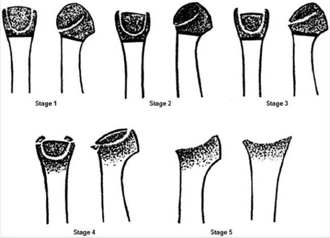
Smillie’s classification for Freiberg’s infarction involves five distinct stages:

Stage 1: Early Fissure and Sclerosis At the onset of Freiberg’s infarction, Stage 1 showcases a fissure in the epiphysis—this is the area of developing bone tissue—and an observable sclerosis between cancellous surfaces. Although symptoms may not be evident, this stage marks the beginning of the condition’s journey.

Stage 2: Absorption and Cartilage Sinking As the condition advances to Stage 2, there’s an absorption of cancellous tissue on the proximal side of the metatarsal head. This absorption prompts the sinking of the articular cartilage dorsally. At this point, patients may begin to experience limited motion and discomfort.

Stage 3: Further Absorption and Bony Projections Progressing to Stage 3, the absorption and sinking of the articular surface intensify. Bony projections emerge both medially and laterally, with the dorsal proximal metatarsal head developing exostosis—a bony outgrowth. This stage underscores the complexity of the condition.

Stage 4: Altered Anatomy and Fractures In Stage 4, the articular surface has sunken significantly, surpassing the point of easy restoration to normal anatomy. It’s important to note that fractures of the medial and lateral projections may occur, accentuating the severity of this stage.

Stage 5: Advanced Arthrosis The final frontier, Stage 5, witnesses the culmination of Freiberg’s infarction. Arthrosis takes center stage, leading to flattening and deformity of the metatarsal head. Interestingly, the plantar aspect retains the original cartilage contour, while the metatarsal shaft thickens and assumes a denser form.

Coughlin & Shurnas classification system for hallux rigidus


The Coughlin and Shurnas classification system is used to categorize different stages of hallux rigidus, a degenerative condition of the big toe joint (first metatarsophalangeal joint) that leads to stiffness and limited motion. This classification system helps surgeons and medical professionals assess the severity of hallux rigidus and determine appropriate treatment strategies.

The Coughlin and Shurnas classification for hallux rigidus consists of four stages:

Stage 0: No arthritic changes are present. The joint has normal or near-normal function, and there is no pain or stiffness.

Stage 1: Mild or early-stage hallux rigidus. Some joint space narrowing and osteophyte formation (bone spurs) are present. There might be mild pain and stiffness with movement.

Stage 2: Moderate hallux rigidus. The joint space is further reduced, and larger osteophytes are present. Joint motion is more restricted, and pain is more pronounced, especially during push-off while walking.

Stage 3: Severe hallux rigidus. The joint space is significantly narrowed or obliterated, and osteophytes are prominent. Joint motion is severely limited, and the patient experiences significant pain and dysfunction, even during walking.

Freiberg’s infarction

Freiberg’s infarction, also known as Freiberg’s disease or osteochondrosis of the metatarsal head, is a condition in which there is a loss of blood supply (avascular necrosis) to the head of the metatarsal bone in the foot. This can cause pain, swelling, and stiffness in the affected foot, and may lead to arthritis or joint deformity over time.

The exact cause of Freiberg’s infarction is not fully understood, but it is believed to be related to repeated trauma or injury to the metatarsal head, which can disrupt the blood supply to the bone. The condition is most commonly seen in adolescent girls and young women, and may be associated with wearing high-heeled shoes or engaging in activities that involve repetitive impact on the foot, such as running or jumping.

Treatment for Freiberg’s infarction typically involves rest, ice, and anti-inflammatory medication to reduce pain and inflammation in the affected foot. Immobilization in a cast or boot may also be recommended to allow the bone to heal. In more severe cases, surgery may be necessary to remove damaged bone or reshape the joint to reduce pain and improve function.

Overall, the prognosis for Freiberg’s infarction depends on the severity of the condition and the effectiveness of the chosen treatment. With appropriate care, many patients are able to manage their symptoms and maintain good foot function over time.

First metatarsophalangeal joint arthrodesis

A first metatarsophalangeal joint (1st MTPJ) arthrodesis is a surgical procedure that involves fusing the bones of the big toe (metatarsal) and the first bone of the toe (phalanx) together to create a solid joint. This procedure is typically performed to treat advanced arthritis, instability, or deformity of the 1st MTPJ.

During the procedure, the surgeon makes an incision on the top of the foot and removes the damaged joint surfaces of the metatarsal and phalanx bones. The bones are then placed in the desired alignment and held in place with screws, pins, or plates until they fuse together. Over time, the bones grow together and form a solid joint, which can help relieve pain and improve the stability and function of the foot.

After the surgery, the foot is immobilized in a cast or boot for several weeks, and physical therapy may be recommended to help regain strength and mobility in the affected foot. While 1st MTPJ arthrodesis can be highly effective in treating certain conditions, it does limit the range of motion in the big toe and may affect the gait or balance of the patient. It is important to discuss the potential risks and benefits of 1st MTPJ arthrodesis with a qualified healthcare professional before undergoing the procedure.

Clinodactyly

Clinodactyly is a medical term describing the curvature of a digit (a finger or toe) in the plane of the palm, most commonly the fifth finger (the “little finger”) towards the adjacent fourth finger (the “ring finger”).

It is a fairly common isolated anomaly which often goes unnoticed, but also occurs in combination with other abnormalities in certain genetic syndromes.[1] The term is from the Ancient Greek κλίνειν klínein ‘to bend’ and δάκτυλος dáktulos ‘digit’.

Webbed toes

Webbed toes, also known as syndactyly, is a condition where two or more toes are fused together by a web of skin and tissue. This can occur in either the feet or the hands, and it is a relatively common condition that affects about 1 in 2,000 to 1 in 3,000 births.

In most cases, webbed toes are not harmful and do not cause any problems with walking or other activities. However, in some cases, the condition may cause difficulty wearing shoes or may be associated with other health problems.

Webbed toes can be treated through surgery, which involves separating the fused digits and reconstructing the skin and soft tissue to create separate toes. The timing of the surgery may depend on the severity of the webbing and whether it is affecting the child’s ability to walk or perform other activities.

It’s important to note that in some cases, webbed toes may be a sign of an underlying genetic condition or syndrome, so it’s important to consult with a healthcare provider if you or your child has webbed toes. They can evaluate the condition and determine whether any further testing or treatment is necessary.

Turf toe

Turf toe is a common injury that occurs when the big toe is forcibly bent upward, causing damage to the ligaments and soft tissues around the joint. It is most commonly seen in athletes who play sports on artificial turf, hence the name “turf toe.”

Symptoms of turf toe include pain, swelling, and stiffness in the affected toe, as well as difficulty walking or bearing weight on the foot. In severe cases, the joint may be dislocated or fractured.

Treatment for turf toe typically involves rest, ice, compression, and elevation (RICE) to reduce pain and swelling. Anti-inflammatory medications may also be recommended to help manage symptoms. In some cases, a stiff-soled shoe or special orthotic device may be prescribed to limit movement of the affected joint and prevent further injury.

Physical therapy may also be recommended to help restore range of motion and strength to the affected foot and prevent future injuries. In rare cases where the joint is severely damaged, surgery may be necessary to repair or reconstruct the ligaments and soft tissues around the joint.

Tailor’s bunion

A tailor’s bunion, also known as a bunionette, is a painful bump that develops on the outside of the foot at the base of the little toe. It is similar to a regular bunion, which develops on the inside of the foot at the base of the big toe.

Tailor’s bunions are usually caused by an inherited structural abnormality in the foot that results in the little toe leaning inward, causing pressure and friction on the outside of the foot. Other factors that can contribute to the development of a tailor’s bunion include wearing tight or poorly fitting shoes, foot injuries, and arthritis.

Symptoms of a tailor’s bunion may include pain, swelling, redness, and a visible bump on the outside of the foot. Treatment for a tailor’s bunion may involve conservative measures such as wearing wider shoes, padding or cushioning the affected area, and taking non-steroidal anti-inflammatory drugs (NSAIDs) to reduce pain and inflammation. In severe cases, surgery may be necessary to correct the underlying structural problem and remove the bony bump.

Prevention of tailor’s bunions involves wearing comfortable, properly fitting shoes that provide enough room for the toes to move freely. Avoiding high heels and shoes with narrow toe boxes can also help reduce the risk of developing a tailor’s bunion. If you experience persistent foot pain or notice any unusual bumps or growths on your feet, it is important to seek medical attention to determine the underlying cause and receive appropriate treatment.

Sesamoid injuries

The sesamoid bones are small bones located under the big toe joint in the foot. They play an important role in weight-bearing and movement of the foot. Sesamoid injuries are a common condition that occurs when the sesamoid bones become injured or inflamed.

There are two types of sesamoid injuries:

  1. Sesamoiditis: This is a condition in which the sesamoid bones become inflamed due to overuse or repetitive stress. Symptoms of sesamoiditis include pain, swelling, and tenderness in the affected area.
  2. Sesamoid Fracture: This is a type of injury that occurs when one or both of the sesamoid bones in the foot are fractured. Symptoms of a sesamoid fracture include severe pain, swelling, and difficulty walking.

To prevent sesamoid injuries, it is important to wear proper footwear that provides adequate cushioning and support, especially if you engage in activities that involve repetitive stress on the foot. In addition, avoid activities that put excessive pressure on the foot, such as jumping and running on hard surfaces.

If you suspect you have a sesamoid injury, it is important to seek medical attention. Treatment for sesamoid injuries may include rest, ice, compression, and elevation (RICE), over-the-counter pain medications, and physical therapy. In severe cases, surgery may be necessary.

Plantar plate tear

The plantar plate is a fibrocartilaginous structure located on the bottom of the foot, which serves to support and stabilize the metatarsophalangeal (MTPJ) joints. A plantar plate tear is a common injury that occurs when the ligamentous fibres of the plantar plate are partially or completely torn.

Plantar plate tears are typically caused by repetitive stress or trauma to the foot, such as from running, jumping, or other high-impact activities. They may also occur as a result of degenerative changes associated with aging or certain medical conditions, such as rheumatoid arthritis.

Symptoms of a plantar plate tear can include pain, swelling, and tenderness in the ball of the foot, particularly when weight-bearing or walking. There may also be a feeling of instability or a “popping” sensation in the affected joint.

Physiotherapy treatment for plantar plate tears may include rest, ice, and immobilisation to reduce pain and inflammation. Manual therapy techniques, such as massage and joint mobilisation, may also be used to improve range of motion and reduce stiffness in the affected area.

Strengthening and stretching exercises can help improve foot and ankle strength and flexibility, and may include exercises to target the plantar fascia and other supporting structures of the foot.

In some cases, custom orthotics or supportive footwear may also be recommended to help relieve pressure on the affected area and prevent further injury.

Surgical intervention may be necessary in cases of severe or chronic plantar plate tears, particularly if conservative treatment is not effective. It is important to consult with a healthcare professional if you suspect you have a plantar plate tear, as early diagnosis and treatment can help prevent further damage and improve overall outcomes.