Category: H

The Herscovici classification for medial malleolar fractures

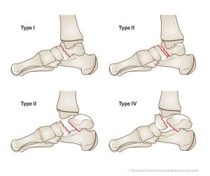
The Herscovici Classification for medial malleolar fractures takes expands from the classifications proposed by Müller et al and Pankovich and Shivaram, with a refined approach that groups fractures into four distinct patterns:

Type-A Fractures: At the simplest end of the spectrum, Type-A fractures involve avulsions of the malleolus tip. While these fractures may seem straightforward, their proper classification aids in tailoring appropriate treatment approaches.

Type-B Fractures: Type-B fractures occur between the tip of the malleolus and the level of the plafond. This pattern introduces a slightly more complex fracture location, necessitating careful consideration during diagnosis and treatment planning.

Type-C Fractures: The Type-C classification involves fractures occurring at the level of the plafond. This represents a pivotal point in the complexity of the fracture pattern and guides healthcare practitioners in devising effective management strategies.

Type-D Fractures: At the upper echelon of complexity, Type-D fractures extend vertically above the level of the plafond. These fractures demand meticulous attention due to their potential implications for treatment and healing.

Hepple MRI staging classification for osteochondral lesions of the talus

The Hepple MRI Staging Classification is a significant framework used to categorize osteochondral lesions of the talus. These lesions involve damage to the cartilage and underlying bone of the ankle joint, often caused by trauma or repetitive stress. The Hepple classification assists in evaluating the severity of such lesions based on MRI findings, aiding in treatment planning and patient management.

The Hepple MRI Staging Classification is divided into four distinct stages:

Stage I: This initial stage is characterized by a subchondral fracture, which appears as a signal change on MRI. The overlying cartilage may remain intact, and there might not be any noticeable separation between the cartilage and the bone. This stage indicates early damage, highlighting the importance of prompt diagnosis and intervention.

Stage II: In Stage II, there is evidence of cartilage separation from the underlying bone, often referred to as a “flap lesion.” This separation can be observed on MRI, and it indicates more significant damage to the osteochondral unit. Timely intervention at this stage can potentially prevent further deterioration.

Stage III: Continuing the progression, Stage III involves a partially detached cartilage fragment within the joint. This fragment is visible on MRI and is indicative of more advanced osteochondral damage. Treatment strategies at this stage may involve addressing the detached fragment to alleviate symptoms and prevent further complications.

Stage IV: The final stage of the Hepple classification represents complete detachment of the cartilage fragment within the joint. The detached fragment can be visualized on MRI and may even displace into the joint space. This stage underscores the urgency of appropriate management, which might include surgical options to restore joint function and prevent long-term consequences.

Hardcastle & Myerson classification for Lisfranc injuries

The Hardcastle and Myerson classification system is used to categorize injuries to the Lisfranc joint complex, which involves the tarsometatarsal joints in the midfoot. This classification system helps surgeons and medical professionals assess the severity of Lisfranc injuries and guide treatment decisions. Lisfranc injuries can range from mild sprains to severe fractures and dislocations.

The Hardcastle and Myerson classification system is divided into three main categories, which are further sub-divided based on the severity of the injury:

Type A: Homolateral Fracture Dislocation

  1. Type A1: Fracture of the base of the second metatarsal.
  2. Type A2: Fracture of the first and second metatarsals.
  3. Type A3: Fracture of all three medial metatarsals.

Type B: Isolated Lisfranc Ligament Injuries

  1. Type B1: Sprain or rupture of the Lisfranc ligament with or without fractures.

Type C: Divergent Dislocation

  1. Type C1: Dislocation of one or two metatarsals.
  2. Type C2: Dislocation of three or more metatarsals.

Each type is associated with varying degrees of instability and severity. Treatment options and outcomes depend on the specific type of Lisfranc injury.

It’s important to note that Lisfranc injuries can be complex and may require careful evaluation by a medical professional, often including imaging studies like X-rays or CT scans. Treatment can range from non-surgical approaches (such as casting or bracing) for less severe injuries to surgical intervention (such as stabilization with screws or plates) for more severe injuries.

If you suspect a Lisfranc injury, it’s crucial to seek prompt medical attention to ensure an accurate diagnosis and appropriate management to prevent potential long-term complications, such as chronic pain and instability in the midfoot.

Surgery considerations for patients with high blood pressure

Patients with high blood pressure (hypertension) require special considerations when undergoing surgery to manage their blood pressure during the perioperative period and minimize the risk of related complications. Here are some important surgery considerations for patients with high blood pressure:

  1. Preoperative evaluation: A thorough preoperative evaluation of the patient’s blood pressure should be conducted to assess the severity of hypertension, identify any associated risk factors or comorbidities, and optimize blood pressure control prior to surgery. This may involve reviewing the patient’s medical history, conducting a physical examination, and performing relevant diagnostic tests, such as blood tests, electrocardiogram (ECG), and echocardiography, as needed.
  2. Blood pressure control optimization: It is important to optimize the patient’s blood pressure control prior to surgery to reduce the risk of perioperative cardiovascular events. This may involve adjusting the patient’s antihypertensive medications, such as diuretics, beta-blockers, calcium channel blockers, and angiotensin-converting enzyme (ACE) inhibitors, to ensure optimal blood pressure management during the perioperative period. Patients with poorly controlled hypertension may require postponement of elective surgery until their blood pressure is better controlled.
  3. Anesthesia considerations: The choice of anesthesia should be carefully considered in patients with hypertension. General anesthesia may be associated with hemodynamic changes, such as changes in blood pressure and heart rate, which can impact patients with hypertension. Therefore, close communication and collaboration between the anesthesiologist and the surgical team are essential to ensure appropriate anesthesia management tailored to the patient’s blood pressure condition. The use of regional anesthesia or monitored anesthesia care may be considered, depending on the type of surgery and the patient’s blood pressure control.
  4. Medication management: Medication management in patients with hypertension should be carefully reviewed and optimized during the perioperative period. This may involve continuing the patient’s regular antihypertensive medications, avoiding triggers, such as nonsteroidal anti-inflammatory drugs (NSAIDs), that may worsen blood pressure control, and having a plan in place for managing blood pressure during and after surgery. In some cases, additional medications, such as short-acting antihypertensive medications, may be used as part of the perioperative management plan.
  5. Intraoperative monitoring: During surgery, close monitoring of the patient’s blood pressure, heart rate, and other relevant hemodynamic parameters should be performed to promptly detect any changes and intervene as needed. This may involve using invasive blood pressure monitoring or continuous noninvasive blood pressure monitoring to ensure optimal blood pressure control during surgery.
  6. Postoperative care: Patients with hypertension may require special postoperative care to manage their blood pressure and prevent cardiovascular complications. This may involve continuing the patient’s regular antihypertensive medications, monitoring blood pressure closely, and providing appropriate pain management that does not negatively impact blood pressure control. Early ambulation, deep breathing exercises, and other preventive measures may also be encouraged to reduce the risk of postoperative complications.
  7. Lifestyle modifications: Patients with hypertension should be encouraged to adopt and maintain healthy lifestyle habits, such as regular exercise, a healthy diet, weight management, stress management, and avoiding tobacco and excessive alcohol consumption. These lifestyle modifications can help improve blood pressure control and overall cardiovascular health, which can have a positive impact on the perioperative management of patients with hypertension.

In conclusion, surgery considerations for patients with high blood pressure require careful evaluation, optimization of blood pressure control, anesthesia considerations, medication management, intraoperative monitoring, postoperative care, and patient education on lifestyle modifications. Collaborative care among the surgical team, anesthesiologist, and other healthcare professionals, along with effective patient education and adherence to blood pressure management protocols, is essential to ensure safe and successful perioperative care for patients with hypertension.

Hawkins classification for talar fractures

The Hawkins classification is a system used to categorize talar fractures, which are fractures of the talus bone in the ankle. The classification was developed by Dr. Herbert Hawkins, an American orthopedic surgeon, in 1970.

The Hawkins classification divides talar fractures into four categories, based on the location and severity of the fracture:

Type I: This is a non-displaced fracture of the talar neck, which is the narrow portion of the talus bone between the body of the talus and the ankle joint. The blood supply to the talus is usually preserved in this type of fracture.

Type II: This is a displaced fracture of the talar neck, which can result in damage to the blood supply to the talus. This type of fracture is considered to be more severe than Type I.

Type III: This is a fracture of the body of the talus, which is the large, rounded portion of the bone that forms the ankle joint. This type of fracture is often associated with significant damage to the blood supply to the talus, and can result in avascular necrosis (death of bone tissue due to loss of blood supply) of the talus.

Type IV: This is a fracture of the posterior process of the talus, which is a small projection on the back of the talus bone. This type of fracture is less common than the other three types, and is generally considered to be less severe.

The Hawkins classification can be useful in guiding treatment decisions for talar fractures, as the severity and location of the fracture can impact the likelihood of complications such as avascular necrosis. Treatment options for talar fractures may include immobilization with a cast or brace, surgery to realign the bones and stabilize the joint, and in some cases, joint replacement surgery.

Overall, the Hawkins classification is a valuable tool for healthcare professionals in assessing and managing talar fractures, and can help improve patient outcomes through more targeted and effective treatment.

How to spot children’s foot problems

As children grow and develop, it is important to pay attention to their foot health to ensure proper growth and function. Here are some tips for parents on how to spot kids’ foot problems:

  1. Pay attention to how your child walks and runs. Look for any limping, toe-walking, or other unusual gait patterns.
  2. Check your child’s shoes regularly to make sure they are not too tight or too loose, and that they are wearing shoes that fit well and provide proper support.
  3. Look for any changes in your child’s foot shape or arches, as these may be signs of developing foot problems.
  4. Pay attention to your child’s complaints of foot pain or discomfort, especially after physical activity or prolonged periods of standing.
  5. Check for any signs of skin irritation, blisters, or calluses on your child’s feet.
  6. If your child has flat feet, is overweight, or participates in high-impact sports, they may be at greater risk for developing foot problems. Be sure to monitor their foot health closely.
  7. Consider taking your child to see a podiatrist if you notice any persistent foot problems or concerns.

By paying attention to your child’s foot health and seeking appropriate care when necessary, you can help ensure they have healthy and happy feet as they grow and develop.

How can I prepare my feet for a marathon?

Preparing your feet for a marathon as a runner is crucial to prevent blisters, calluses, and other foot problems that can hinder your performance. Here are some tips to help you prepare your feet for a marathon:

  1. Choose the right shoes: Make sure you have comfortable and supportive running shoes that fit well. Choose shoes that are designed for long-distance running and that have enough room in the toe box.
  2. Break in your shoes: Avoid wearing brand new shoes on race day. Break in your shoes by wearing them for shorter runs in the weeks leading up to the marathon.
  3. Wear moisture-wicking socks: Choose socks made of moisture-wicking material to keep your feet dry and prevent blisters.
  4. Apply anti-chafing products: Apply anti-chafing products, such as petroleum jelly or body glide, to areas of the feet that are prone to rubbing or blistering.
  5. Trim your toenails: Trim your toenails before race day to prevent ingrown toenails and other foot problems.
  6. Strengthen your feet: Incorporate exercises to strengthen your feet, such as toe curls and heel raises, into your training routine.
  7. Stretch your feet: Stretch your feet before and after each run to help prevent cramping and improve flexibility.

By following these tips, you can help prevent foot problems and ensure that your feet are ready for the marathon. It is also important to listen to your body and seek medical attention if you experience pain or discomfort in your feet.

I am concerned with my child’s feet. What should I do?

If you are concerned about your child’s feet, there are several things you can do:

  1. Observe their feet: Take a look at your child’s feet and observe their walking and standing posture. Look for any unusual features, such as flat feet, high arches, or toe abnormalities.
  2. Talk to your child: Ask your child if they are experiencing any pain or discomfort in their feet, and whether they are having any difficulty with walking or participating in sports.
  3. Consult a paediatrician or podiatrist: If you have any concerns, schedule an appointment with your child’s paediatrician or a podiatrist. They can evaluate your child’s feet and provide recommendations for treatment or further evaluation if necessary.
  4. Provide proper footwear: Make sure your child wears properly fitting shoes that provide adequate support and protection for their feet. Avoid shoes with high heels or those that are too tight.
  5. Encourage physical activity: Regular physical activity can help promote foot health and development in children.

By taking these steps, you can help ensure your child’s feet remain healthy and address any concerns early on.

How do I improve my foot health?

There are several ways to improve your foot health, including:

  1. Wear proper shoes: Choose shoes that fit well and provide proper support for your feet. Avoid wearing high heels or shoes that are too tight, which can lead to foot problems.
  2. Practice good foot hygiene: Wash your feet daily with soap and water and dry them thoroughly, paying special attention to the area between your toes. Apply moisturiser to prevent dryness and cracking.
  3. Exercise regularly: Regular physical activity can help improve circulation and strengthen your feet and ankles.
  4. Stretch your feet and ankles: Stretching can help improve flexibility and reduce the risk of injury. Try stretching exercises such as toe raises, ankle circles, and calf stretches.
  5. Maintain a healthy weight: Excess weight can put extra strain on your feet and lead to foot problems.
  6. Check your feet regularly: Inspect your feet regularly for any signs of injury or infection, such as cuts, blisters, or redness.
  7. Seek medical attention when necessary: If you experience persistent foot pain, swelling, or other symptoms, see a healthcare professional for diagnosis and treatment.

By following these tips, you can help improve your foot health and reduce the risk of foot problems.

How do I get a piece of broken glass out of my foot?

If you have a piece of broken glass in your foot, it is important to remove it as soon as possible to prevent infection and further injury. Here are the steps to follow:

  1. Clean the area: Wash the area with soap and water and gently pat it dry with a clean towel.
  2. Inspect the wound: Use a magnifying glass if necessary to locate the piece of glass.
  3. Sterilise the area: Use rubbing alcohol or another antiseptic solution to sterilise the area around the wound.
  4. Use tweezers: Use a pair of sterile tweezers to grasp the piece of glass firmly at the base and gently pull it out in the same direction it entered.
  5. Clean the wound: Once the glass is removed, wash the wound again with soap and water and apply an antiseptic ointment.
  6. Cover the wound: Cover the wound with a sterile adhesive bandage or sterile gauze and secure it in place with medical tape.

If you are unable to remove the glass or if you experience any signs of infection such as redness, swelling, or fever, seek medical attention from a healthcare professional.