Category: Hindfoot

Posterior tibial tendon dysfunction

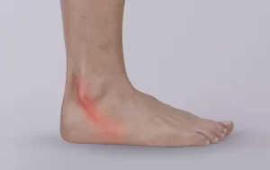
Posterior tibial tendon dysfunction (PTTD) is a condition that occurs when the posterior tibial tendon, which runs along the inside of the ankle and foot, becomes damaged or inflamed. The posterior tibial tendon is responsible for supporting the arch of the foot and helping to maintain proper alignment and stability.

PTTD is often caused by overuse or repetitive stress on the posterior tibial tendon, which can lead to small tears or degeneration of the tendon. Other factors that may contribute to PTTD include obesity, flat feet, high-impact activities, and certain medical conditions such as rheumatoid arthritis.

Symptoms of PTTD may include pain and swelling along the inside of the ankle and foot, particularly during and after physical activity, as well as a gradual flattening of the arch of the foot and a shifting of the heel bone outward.

Treatment for PTTD typically involves a combination of non-surgical interventions such as rest, ice, compression, elevation, and physical therapy to help reduce pain and inflammation and improve strength and flexibility in the affected foot and ankle. Orthotic devices such as arch supports or braces may also be recommended to help support the foot and relieve stress on the posterior tibial tendon.

In more severe cases of PTTD, surgical intervention may be necessary to repair or reconstruct the damaged tendon, restore proper alignment of the foot, and improve overall foot and ankle function. It is important to consult with a healthcare professional to determine the most appropriate treatment plan for your specific condition and individual health needs.

Plantar fasciitis – what happens at a cellular level?

Plantar fasciitis is a condition that involves inflammation and damage to the plantar fascia, which is a thick band of connective tissue that runs along the bottom of the foot. At a cellular level, plantar fasciitis involves a complex interplay of inflammatory mediators and cellular responses.

One of the key cellular processes involved in plantar fasciitis is the release of inflammatory mediators, such as cytokines, from damaged tissue cells. These mediators trigger a cascade of cellular responses, including the activation of immune cells, the release of more inflammatory mediators, and the recruitment of additional cells to the site of injury.

As inflammation persists, it can lead to the breakdown of extracellular matrix components, including collagen and elastin fibres, which are important structural components of the plantar fascia. This breakdown can further weaken the tissue and contribute to ongoing inflammation and pain.

Over time, chronic inflammation and tissue damage can lead to the formation of scar tissue and the thickening of the plantar fascia, which can further exacerbate symptoms of plantar fasciitis.

Interventions for plantar fasciitis aim to address these underlying cellular processes by reducing inflammation, promoting tissue healing, and restoring normal biomechanical function to the foot and ankle. By addressing the cellular mechanisms involved in plantar fasciitis, treatment can help alleviate symptoms, prevent further damage, and promote long-term recovery.

Plantar fasciitis

Plantar fasciitis is a common condition that causes pain in the heel and bottom of the foot. It is caused by inflammation of the plantar fascia, a thick band of tissue that runs along the bottom of the foot and connects the heel bone to the toes.

Symptoms of plantar fasciitis include pain and tenderness in the heel or arch of the foot, particularly upon waking in the morning or after prolonged periods of standing or walking. The pain may be described as sharp or stabbing, and may worsen over time if left untreated.

Interventions for plantar fasciitis may include:

  1. Stretching exercises: Specific stretches for the calf, Achilles tendon, and plantar fascia can help improve flexibility and reduce tension in the affected area.
  2. Strengthening exercises: Strengthening exercises for the foot and ankle can help improve stability and support the plantar fascia.
  3. Manual therapy: Massage, joint mobilisation, and other hands-on techniques can help reduce pain and improve mobility.
  4. Footwear advice: Proper footwear can help reduce stress on the plantar fascia and prevent further injury.
  5. Other Modalities: The use of modalities such as ice, heat, ultrasound, or electrical stimulation can help reduce pain and inflammation.
  6. Activity modification: Modifying or avoiding activities that exacerbate the symptoms of plantar fasciitis can help reduce pain and promote healing.

It is important to consult with a healthcare professional if you are experiencing symptoms of plantar fasciitis, as early diagnosis and treatment can help prevent further damage and improve overall outcomes. With appropriate physiotherapy care, most individuals with plantar fasciitis can expect to make a full recovery and return to their normal activities.

Peroneal tendon instability

Peroneal tendon instability is a condition in which the peroneal tendons, which run along the outside of the ankle and foot, are not held in place properly and are prone to dislocation or subluxation (partial dislocation). This can cause pain, weakness, and instability in the ankle and foot.

Peroneal tendon instability is often caused by a combination of factors, including ankle sprains, chronic ankle instability, and anatomical abnormalities that affect the stability of the peroneal tendons.

Symptoms of peroneal tendon instability can include pain and tenderness along the outside of the ankle and foot, a feeling of snapping or popping in the ankle, weakness and instability, and swelling and stiffness.

Treatment for peroneal tendon instability may include non-surgical options such as physical therapy to strengthen the ankle and foot muscles, bracing or taping to provide additional support, and modification of activities to avoid aggravating the condition.

In some cases, surgical intervention may be necessary to repair or reconstruct the damaged peroneal tendons, particularly if non-surgical treatments are not effective or the instability is severe.

It is important to consult with a healthcare professional if you are experiencing symptoms of peroneal tendon instability, as early diagnosis and treatment can help prevent further damage and improve overall outcomes. With appropriate care, most individuals with peroneal tendon instability can expect to make a full recovery and return to their normal activities.

Peroneal tendon injuries

The peroneal tendons are two long tendons that run along the outside of the ankle and foot, connecting the muscles of the lower leg to the bones of the foot. Peroneal tendon injuries can occur due to a variety of factors, including overuse, trauma, or anatomical abnormalities.

Symptoms of peroneal tendon injuries can include pain, swelling, and tenderness along the outside of the ankle and foot, as well as weakness and instability. In some cases, individuals may also experience a popping or snapping sensation in the ankle.

Treatment for peroneal tendon injuries can vary depending on the severity of the injury. Mild injuries may be treated with rest, ice, compression, and elevation, as well as non-steroidal anti-inflammatory drugs (NSAIDs) to manage pain and inflammation. Physical therapy may also be recommended to help strengthen the muscles and improve flexibility.

In more severe cases, immobilisation in a cast or brace may be necessary to allow the tendon to heal. In some cases, surgery may be necessary to repair or reconstruct the damaged tendon.

It is important to consult with a healthcare professional if you are experiencing symptoms of a peroneal tendon injury, as early diagnosis and treatment can help prevent further damage and improve overall outcomes. With appropriate care, most individuals with peroneal tendon injuries can expect to make a full recovery and return to their normal activities.

Heel pain

Heel pain is a common condition that can be caused by a variety of factors. The most common cause of heel pain is plantar fasciitis, which is an inflammation of the plantar fascia, a thick band of tissue that runs along the bottom of the foot from the heel to the toes.

Other causes of heel pain may include:

  • Achilles tendinitis: inflammation of the Achilles tendon that connects the calf muscle to the heel bone
  • Heel bursitis: inflammation of the fluid-filled sacs that cushion the heel bone
  • Heel spurs: bony growths that develop on the heel bone due to long-term plantar fasciitis
  • Stress fractures: tiny cracks in the bones of the foot that can cause heel pain and swelling
  • Tarsal tunnel syndrome: compression of the nerve that runs along the inside of the ankle and into the foot

Symptoms of heel pain may include:

  • Pain or tenderness in the heel or arch of the foot
  • Stiffness or swelling in the heel or foot
  • Difficulty standing or walking for long periods of time
  • Pain that is worse in the morning or after prolonged periods of rest
  • Pain that improves with activity but worsens after exercise

Treatment for heel pain depends on the underlying cause. In most cases, conservative measures such as rest, ice, stretching exercises, and over-the-counter pain medications may be sufficient. In more severe cases, a podiatrist may recommend custom orthotics, physical therapy, or other interventions to help relieve pain and improve function.

If you are experiencing heel pain, it is important to seek medical attention for an accurate diagnosis and appropriate treatment. Early intervention can help prevent the condition from worsening and reduce the risk of long-term complications.

Haglund’s deformity

Haglund’s deformity, also known as “pump bump,” is a bony enlargement on the back of the heel bone. It is typically caused by pressure and friction from shoes rubbing against the back of the heel. The symptoms of Haglund’s deformity include pain, swelling, redness, and a visible bump on the back of the heel.

Treatment for Haglund’s deformity typically involves conservative measures such as wearing shoes with a soft back, using heel pads or lifts to reduce pressure on the heel, and taking anti-inflammatory medications to reduce pain and swelling. Physical therapy exercises and stretching may also help to improve flexibility and reduce symptoms.

In some cases, surgery may be necessary to remove the bony protrusion and relieve symptoms. This may be recommended if conservative measures do not provide relief, or if the deformity is severe and causing significant pain and disability.

It is important to seek medical attention if you are experiencing symptoms of Haglund’s deformity, as early diagnosis and treatment can help to prevent the condition from worsening and improve overall outcomes.

Diffuse idiopathic skeletal hyperostosis (DISH)

Diffuse Idiopathic Skeletal Hyperostosis (DISH) is a condition in which there is abnormal growth of bone along the sides of the vertebrae of the spine, as well as in other areas of the body, such as the hips, shoulders, knees, and heels. The cause of DISH is unknown, but it is more commonly seen in older adults, particularly those who are overweight or have diabetes.

The excessive growth of bone in DISH can lead to stiffness and pain in the affected areas, as well as difficulty moving. In some cases, DISH can also cause pressure on nearby nerves, leading to additional symptoms such as numbness or tingling.

Diagnosis of DISH is typically made through imaging tests such as X-rays or CT scans, which can show the bony growths. Treatment for DISH usually involves managing symptoms, such as pain and stiffness, with medications or physical therapy. Surgery may be considered in severe cases, particularly if there is nerve compression or other complications.

It is important for people with DISH to maintain a healthy weight, exercise regularly, and manage any underlying health conditions, such as diabetes, to help prevent the progression of the condition. Regular follow-up with a healthcare provider is also recommended to monitor any changes and adjust treatment as necessary.

Calcaneal fracture (heel fracture)

A calcaneal fracture is a break in the heel bone, also known as the calcaneus. This type of fracture is typically caused by a high-energy injury, such as a fall from a height or a car accident. It can also occur as a result of repetitive stress, particularly in athletes who engage in high-impact activities like running and jumping.

Symptoms of a calcaneal fracture include:

  1. Severe pain in the heel, particularly when bearing weight
  2. Swelling and bruising around the heel
  3. Inability to walk or put weight on the affected foot
  4. Tenderness when touching the heel
  5. Deformity or flattening of the heel

Treatment for a calcaneal fracture depends on the severity of the injury. In some cases, nonsurgical treatment, such as immobilisation in a cast or boot, may be sufficient to allow the bone to heal. However, in more severe cases, surgery may be required to realign and stabilise the bone.

Recovery from a calcaneal fracture can be a lengthy process, often taking several months for the bone to fully heal. During this time, physical therapy may be necessary to help regain strength and mobility in the affected foot. It is also important to avoid weight-bearing activities until the bone has fully healed, to prevent re-injury or complications.

Calcaneal apophysitis (Sever’s disease)

Calcaneal apophysitis, also known as Sever’s disease, is a common cause of heel pain in children and adolescents. It is an overuse injury that occurs when there is inflammation of the growth plate in the heel bone, where the Achilles tendon attaches. It typically affects children between the ages of 8 and 15, particularly those who are involved in sports that involve running and jumping.

Symptoms of calcaneal apophysitis include:

  1. Pain in the heel, particularly with activity
  2. Swelling and tenderness in the heel
  3. Difficulty walking or running
  4. Stiffness in the heel upon waking up in the morning

Treatment for calcaneal apophysitis typically involves rest, ice, compression, and elevation (RICE). Anti-inflammatory medications, such as ibuprofen, may also be used to reduce pain and inflammation. In some cases, a heel cup or other supportive device may be recommended to help cushion the heel and reduce stress on the growth plate.

It is important for children and adolescents with calcaneal apophysitis to avoid activities that aggravate the condition until the symptoms have completely resolved. In most cases, symptoms will resolve within a few weeks to a few months with proper treatment and rest. However, if left untreated, calcaneal apophysitis can lead to chronic heel pain and other complications.