Author: Steve

Valleix’s sign

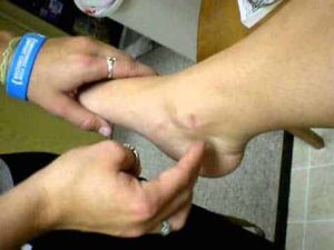
Valleix’s sign is another diagnostic test that can be used to assess nerve function in the peripheral nervous system. It involves applying pressure or pinching along the course of a nerve to identify areas of tenderness or pain.

In the case of tarsal tunnel syndrome, the Valleix sign can be used to identify areas of nerve irritation or damage along the course of the posterior tibial nerve. The healthcare provider will apply pressure or pinch along the nerve pathway, starting at the ankle and moving up the leg, while asking the patient if they feel any pain or discomfort.

If the patient experiences pain or tenderness along the course of the posterior tibial nerve, this can be an indication of nerve irritation or damage. However, like Tinel’s sign, the Valleix sign is not always definitive, and additional diagnostic tests may be needed to confirm the diagnosis.

In general, a combination of diagnostic tests and a thorough medical history and physical exam are used to diagnose tarsal tunnel syndrome. Treatment may involve rest, ice, physical therapy, and other conservative measures, and in some cases, surgery may be necessary to relieve pressure on the affected nerve.

Tinel’s sign

Tinel’s sign is a diagnostic test used to assess nerve function, particularly in the peripheral nervous system. Tarsal tunnel syndrome is a condition in which the posterior tibial nerve, which runs through the tarsal tunnel in the ankle, becomes compressed or damaged, resulting in pain, numbness, or tingling in the foot.

To perform Tinel’s sign for tarsal tunnel syndrome, a healthcare provider will tap gently on the affected area near the posterior tibial nerve. If the patient experiences a tingling sensation or a shock-like sensation that radiates into the foot, this can be an indication of nerve irritation or damage.

While Tinel’s sign can be useful in diagnosing tarsal tunnel syndrome, it is not always definitive, and other diagnostic tests, such as nerve conduction studies, may also be needed to confirm the diagnosis. Treatment for tarsal tunnel syndrome typically involves conservative measures such as rest, ice, and physical therapy, but in some cases, surgery may be necessary to relieve pressure on the affected nerve.

Scintigraphy (bone scan) for feet

Scintigraphy, also known as a bone scan, is a medical imaging technique that uses a small amount of radioactive material to produce images of bones in the body. It is often used to evaluate the bones of the feet and to diagnose and monitor conditions such as fractures, infections, arthritis, and tumors.

During a scintigraphy procedure, a small amount of a radioactive tracer material is injected into a vein in the patient’s arm. The tracer material then travels through the bloodstream and accumulates in areas of bone that are undergoing active changes or have an increased blood supply. A special camera then captures images of the foot, showing areas of the bone that have taken up the tracer.

Scintigraphy is a safe and non-invasive procedure that typically takes between 1-3 hours to complete. Patients may be asked to lie still on a table during the procedure. The amount of radiation exposure from a bone scan is generally considered to be very low and poses little risk to the patient.

Scintigraphy for the feet can help identify bone abnormalities that may not be visible on X-rays or other imaging tests. It can also provide valuable information about the extent and severity of conditions affecting the bones of the feet, which can help guide treatment decisions.

Your doctor may recommend scintigraphy for the feet if you have symptoms such as foot pain, swelling, or limited mobility, or if they suspect a bone abnormality that cannot be identified through other imaging techniques.

SPECT-CT for feet

SPECT-CT (Single Photon Emission Computed Tomography-Computed Tomography) is a medical imaging technique that combines two imaging modalities, SPECT and CT, to create a three-dimensional image of the body. It is often used to diagnose and evaluate conditions in the feet, such as fractures, infections, and tumors.

SPECT uses a small amount of a radioactive substance called a tracer, which is injected into the patient’s bloodstream. The tracer emits gamma rays, which are detected by a specialized camera to create images of the foot’s internal structures. CT uses X-rays to create detailed images of the bones and soft tissues in the foot. Combining the two modalities allows for more accurate and detailed images than either technique alone.

SPECT-CT for the feet is a non-invasive procedure that typically takes between 30-60 minutes to complete. Patients are asked to lie down on a table and remain still while the images are being taken. The procedure is generally considered safe, but as with any medical imaging test, there is a small risk of exposure to radiation.

SPECT-CT for feet can help diagnose a variety of conditions and can aid in treatment planning by providing detailed information about the location and extent of abnormalities in the feet. Your doctor will determine if this imaging test is appropriate for your specific situation.

Skew foot

Skewfoot, also known as oblique foot or serpentine foot, is a rare foot deformity that can be present at birth or develop over time. It is characterized by an abnormal position of the heel bone (calcaneus) and the midfoot bones, resulting in a foot that is twisted and tilted inwards.

The exact cause of skewfoot is not fully understood, but it is believed to be related to abnormal development of the bones and joints in the foot. The condition can also be associated with other foot and leg problems, such as flat feet, high arches, and hip dysplasia.

The symptoms of skewfoot can include pain, stiffness, and difficulty walking or standing for long periods of time. Treatment options may include wearing orthotics or special shoes, physical therapy to improve foot alignment and range of motion, or surgery to correct the deformity in severe cases.

If you suspect you have skewfoot, it is recommended that you consult with a podiatrist or orthopedic surgeon who specializes in foot and ankle conditions for a proper diagnosis and treatment plan.

Polymetatarsia

Polymetatarsia is a rare condition in which there are more than five metatarsal bones in the forefoot. The most common type of polymetatarsia involves an additional metatarsal bone between the fourth and fifth metatarsals, which is called a “supernumerary metatarsal.” This extra bone can cause pain, discomfort, and difficulty with shoe fitting.

Polymetatarsia may be congenital (present at birth) or acquired (due to injury or disease). It can be diagnosed through physical examination, X-rays, and other imaging studies. Treatment options include wearing custom orthotics or padding to relieve pressure on the foot, modifying shoes to accommodate the extra bone, or surgical removal of the supernumerary metatarsal. The best treatment option depends on the individual’s symptoms and the severity of the condition.

Progressive collapsing flatfoot deformity – Classification system

Progressive collapsing flatfoot deformity can be classified according to the severity of the deformity, as well as the underlying cause. There are several classification systems used by healthcare providers to describe the different stages of the condition. Here are two common classification systems:

  1. Johnson and Strom’s classification system:
  • Stage I: The foot is flat when bearing weight, but the arch returns when the foot is lifted off the ground.
  • Stage II: The arch does not return when the foot is lifted off the ground, but the foot is still flexible.
  • Stage III: The foot is rigid, and there is significant arthritis and deformity.
  1. Myerson’s classification system:
  • Stage I: The hindfoot is in a valgus position (tilted outward) and the arch is slightly flattened.
  • Stage II: The hindfoot is in a valgus position, the arch is more severely flattened, and there is evidence of instability in the ankle joint.
  • Stage III: The hindfoot is in a valgus position, the arch is severely flattened, and there is significant ankle joint arthritis.

The classification system used will depend on the healthcare provider’s preference and the specific needs of the patient.

Progressive collapsing flatfoot deformity

Progressive collapsing flatfoot deformity is a condition in which the arch of the foot gradually collapses over time, causing the foot to become flatter and longer. This condition is typically caused by a combination of factors, including genetics, obesity, injury, and age.

As the arch of the foot collapses, the foot becomes less stable, which can lead to pain and discomfort in the foot, ankle, and lower leg. Other symptoms may include swelling, stiffness, and aching in the foot and ankle.

Treatment for progressive collapsing flatfoot deformity may include a combination of non-surgical and surgical approaches. Non-surgical treatment may include rest, ice, compression, elevation, physical therapy, and the use of supportive footwear and orthotics. Surgical treatment may be recommended in cases where non-surgical methods are not effective, and may involve procedures such as tendon transfers, bone realignment, or fusion.

If you are experiencing symptoms of progressive collapsing flatfoot deformity, it is important to consult with a healthcare provider for proper diagnosis and treatment recommendations.

Merton Root

Merton Root (1915-1995) was an American podiatrist who made significant contributions to the field of podiatry, particularly in the area of biomechanics. He developed a systematic approach to the examination, diagnosis, and treatment of foot and ankle disorders that came to be known as the “Root method.”

The Root method emphasizes the importance of understanding the relationship between the foot’s structure and its function, and how abnormalities in one can affect the other. Root believed that proper diagnosis and treatment of foot disorders required a comprehensive understanding of these relationships.

Root was also a co-founder of the American Academy of Podiatric Sports Medicine and was instrumental in establishing podiatry as a recognized medical specialty in the United States. His contributions to the field of podiatry continue to be recognized and studied by podiatrists today.

Polydactyly

Polydactyly is a genetic condition characterized by the presence of extra fingers or toes on a person’s hands or feet. Instead of the typical five digits on each hand or foot, a person with polydactyly may have six, seven, or even more digits. The extra digits can be fully formed and functional or they may be small, non-functional nubs.

Polydactyly can be inherited in an autosomal dominant pattern, meaning that a child has a 50% chance of inheriting the condition if one parent carries the gene. In some cases, however, polydactyly can occur spontaneously as a random mutation.

Polydactyly is usually not a serious medical condition, and many people with the condition are able to live normal, healthy lives without any problems. In some cases, however, surgery may be necessary to remove the extra digits if they are causing problems with mobility or other issues.