Author: Steve

Forestier’s disease

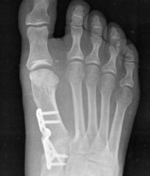
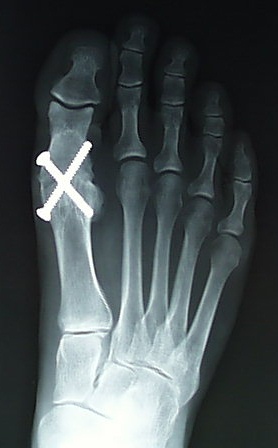
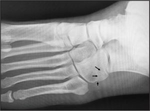
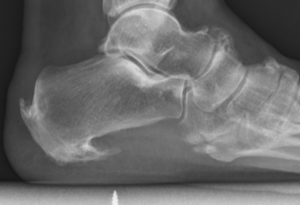
Forestier’s disease can affect various parts of the body, including the spine, pelvis, hip, and knee, but it is less common for it to affect the foot. However, in rare cases, Forestier’s disease can cause abnormal bone growth in the foot, leading to stiffness, pain, and difficulty with movement.

The symptoms of Forestier’s disease in the foot may include:

  • Stiffness and limited range of motion in the affected joint(s)
  • Pain that worsens with activity
  • Swelling or tenderness around the affected area
  • Deformity or abnormal bone growth in the foot

Treatment for Forestier’s disease in the foot may include nonsteroidal anti-inflammatory drugs (NSAIDs) for pain and inflammation, physical therapy to improve range of motion and mobility, and in severe cases, surgery to remove or reshape the abnormal bone growth. It’s important to consult with a healthcare provider for proper diagnosis and treatment of Forestier’s disease in the foot.

Lapidus procedure

The Lapidus procedure is a surgical procedure used to treat hallux valgus, a condition in which the big toe drifts away from the midline of the foot, causing a bunion. It involves fusing the first metatarsal bone to the medial cuneiform bone in the midfoot to correct the alignment of the bones and reduce the deformity. This procedure is typically reserved for cases of severe hallux valgus or for patients who have not responded to more conservative treatments.

During the procedure, the surgeon makes an incision on the top of the foot and removes a small piece of bone from the base of the first metatarsal. The metatarsal bone is then repositioned and fixed in place with screws or a plate to hold it in the desired alignment. Over time, the bones grow together and form a solid fusion, which helps to stabilize the midfoot and reduce the severity of the bunion.

After the surgery, the patient may need to wear a cast or brace for several weeks to protect the foot and allow the bones to fuse together. Physical therapy may also be recommended to help improve strength, flexibility, and range of motion in the affected foot. While the Lapidus procedure can be highly effective in correcting hallux valgus, it does require a period of immobilization and recovery, and may have some potential risks and complications, such as non-union, nerve injury, or infection. It is important to discuss the potential risks and benefits of the Lapidus procedure with a qualified healthcare professional before undergoing the procedure.

Freiberg’s infarction

Freiberg’s infarction, also known as Freiberg’s disease or osteochondrosis of the metatarsal head, is a condition in which there is a loss of blood supply (avascular necrosis) to the head of the metatarsal bone in the foot. This can cause pain, swelling, and stiffness in the affected foot, and may lead to arthritis or joint deformity over time.

The exact cause of Freiberg’s infarction is not fully understood, but it is believed to be related to repeated trauma or injury to the metatarsal head, which can disrupt the blood supply to the bone. The condition is most commonly seen in adolescent girls and young women, and may be associated with wearing high-heeled shoes or engaging in activities that involve repetitive impact on the foot, such as running or jumping.

Treatment for Freiberg’s infarction typically involves rest, ice, and anti-inflammatory medication to reduce pain and inflammation in the affected foot. Immobilization in a cast or boot may also be recommended to allow the bone to heal. In more severe cases, surgery may be necessary to remove damaged bone or reshape the joint to reduce pain and improve function.

Overall, the prognosis for Freiberg’s infarction depends on the severity of the condition and the effectiveness of the chosen treatment. With appropriate care, many patients are able to manage their symptoms and maintain good foot function over time.

First metatarsophalangeal joint arthrodesis

A first metatarsophalangeal joint (1st MTPJ) arthrodesis is a surgical procedure that involves fusing the bones of the big toe (metatarsal) and the first bone of the toe (phalanx) together to create a solid joint. This procedure is typically performed to treat advanced arthritis, instability, or deformity of the 1st MTPJ.

During the procedure, the surgeon makes an incision on the top of the foot and removes the damaged joint surfaces of the metatarsal and phalanx bones. The bones are then placed in the desired alignment and held in place with screws, pins, or plates until they fuse together. Over time, the bones grow together and form a solid joint, which can help relieve pain and improve the stability and function of the foot.

After the surgery, the foot is immobilized in a cast or boot for several weeks, and physical therapy may be recommended to help regain strength and mobility in the affected foot. While 1st MTPJ arthrodesis can be highly effective in treating certain conditions, it does limit the range of motion in the big toe and may affect the gait or balance of the patient. It is important to discuss the potential risks and benefits of 1st MTPJ arthrodesis with a qualified healthcare professional before undergoing the procedure.

Subtalar joint arthrodesis

A subtalar joint arthrodesis is a surgical procedure in which the subtalar joint, which is located between the heel bone (calcaneus) and the ankle bone (talus), is fused together to create a single, solid bone. This procedure is typically performed to treat conditions such as severe arthritis, instability, or deformity of the subtalar joint.

During the procedure, the surgeon makes an incision on the side of the foot and removes the damaged cartilage from the subtalar joint. The bones are then positioned in the desired alignment and held in place with screws or other hardware. Over time, the bones grow together and fuse into a single, solid bone.

Subtalar joint arthrodesis is typically performed under general anesthesia and requires a period of immobilization in a cast or brace. Physical therapy is also recommended to help regain strength and mobility in the affected foot. While this procedure can be highly effective in treating certain conditions, it does limit the range of motion in the foot and ankle and may lead to an increased risk of arthritis in adjacent joints over time. It is important to discuss the potential risks and benefits of subtalar joint arthrodesis with a qualified healthcare professional before undergoing the procedure.

Instep fasciotomy (plantar fasciitis surgery)

An instep plantar fasciotomy is a surgical procedure that involves cutting a portion of the plantar fascia ligament in the foot to relieve tension and pain. The plantar fascia is a thick band of tissue that runs along the bottom of the foot, connecting the heel bone to the toes. When this tissue becomes inflamed or irritated, it can cause a condition called plantar fasciitis, which is characterized by pain and stiffness in the heel and sole of the foot.

During an instep plantar fasciotomy, the surgeon makes a small incision on the top of the foot, near the base of the toes, and cuts a portion of the plantar fascia ligament. This reduces tension on the ligament and relieves pressure on the affected area. The incision is typically closed with stitches, and the foot is immobilized in a cast or boot for several weeks to allow for proper healing.

While instep plantar fasciotomy can be an effective treatment for plantar fasciitis, it is typically reserved for cases that have not responded to non-surgical treatments, such as rest, stretching, and physical therapy. As with any surgical procedure, there are risks and potential complications associated with instep plantar fasciotomy, and it is important to discuss these with a qualified healthcare professional before undergoing the procedure.

Gastrocnemius recession (calf release)

A gastrocnemius recession, also known as gastrocnemius lengthening or calf muscle release, is a surgical procedure in which the gastrocnemius muscle is partially cut to relieve tension and tightness in the calf. This procedure is typically performed to treat a condition called equinus deformity, which is characterized by limited ankle dorsiflexion (the ability to pull the foot upward toward the shin).

During the procedure, a small incision is made in the skin over the gastrocnemius muscle, and the muscle is partially cut or released from its attachment to the heel bone (calcaneus). This allows the muscle to stretch more easily and reduces tension on the Achilles tendon and plantar fascia.

Gastrocnemius recession is often performed in combination with other procedures, such as Achilles tendon lengthening or plantar fascia release, to address the underlying cause of equinus deformity. Recovery from the procedure typically involves a period of immobilization and physical therapy to regain strength and flexibility in the affected leg. As with any surgery, there are risks and potential complications associated with gastrocnemius recession, and it is important to discuss these with a qualified healthcare professional before undergoing the procedure.

Clinodactyly

Clinodactyly is a medical term describing the curvature of a digit (a finger or toe) in the plane of the palm, most commonly the fifth finger (the “little finger”) towards the adjacent fourth finger (the “ring finger”).

It is a fairly common isolated anomaly which often goes unnoticed, but also occurs in combination with other abnormalities in certain genetic syndromes.[1] The term is from the Ancient Greek κλίνειν klínein ‘to bend’ and δάκτυλος dáktulos ‘digit’.

Iontophoresis

Iontophoresis is a non-invasive medical treatment that uses a low-level electrical current to deliver medication through the skin. It is commonly used to treat hyperhidrosis, a medical condition characterized by excessive sweating, but can also be used for other purposes, such as pain management and drug delivery.

During the treatment, electrodes are placed on the skin, and a mild electrical current is applied to the affected area. This current creates an electric field that allows charged ions, such as medication molecules, to move through the skin and into the underlying tissues.

Iontophoresis is a safe and effective treatment that can be used in a variety of medical settings, including physical therapy clinics, hospitals, and doctor’s offices. However, it is important to note that not all medications can be delivered using iontophoresis, and the treatment may not be suitable for everyone. It is important to consult with a medical professional to determine if iontophoresis is an appropriate treatment option for your specific condition.

Nutcracker fracture

A nutcracker fracture is a type of foot fracture that occurs when the cuboid bone, located on the outer side of the foot, is crushed between the heel bone and the fourth and fifth metatarsal bones.

This type of fracture can occur as a result of a sudden injury, such as a fall or a direct blow to the foot, or from overuse or repetitive stress on the foot. Symptoms of a nutcracker fracture of the cuboid may include pain, swelling, bruising, and difficulty bearing weight on the affected foot.

Diagnosis is typically made through a physical exam and imaging tests, such as X-rays or CT scans. Treatment for a nutcracker fracture of the cuboid may depend on the severity of the fracture, but generally involves immobilizing the affected foot with a cast or brace and allowing time for the bone to heal. In some cases, surgery may be necessary to realign the fractured bone and stabilize the foot. Physical therapy may also be recommended to help restore range of motion and strength to the affected foot.