Category: Classification systems

Torg classification for fifth metatarsal fractures

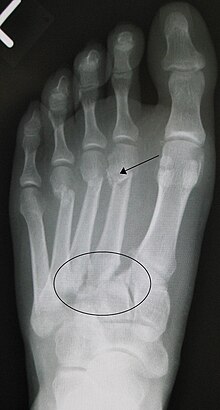
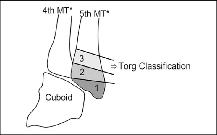
The Torg Classification, developed by Dr. J.W. Torg, is a classification system for fractures of the fifth metatarsal bone. This classification is primarily used to categorize fractures in athletes and guide treatment decisions based on the location and characteristics of the fracture.

The Torg Classification divides fifth metatarsal fractures into three zones:

Zone 1: Avulsion Fractures

  • Involves an avulsion of the tuberosity of the fifth metatarsal due to pull of the peroneus brevis tendon.
  • Often referred to as a “dancer’s fracture” or “pseudo-Jones fracture.”
  • Typically has a good prognosis and usually heals well with conservative treatment.

Zone 2: Jones Fractures

  • Occurs at the metaphyseal-diaphyseal junction of the fifth metatarsal, approximately 1.5 to 3 cm distal to the tuberosity.
  • These fractures are more prone to delayed or non-union due to limited blood supply in this area.
  • Often requires more aggressive treatment, including immobilization and sometimes surgical intervention.

Zone 3: Diaphyseal Fractures

  • Involves fractures of the diaphysis (shaft) of the fifth metatarsal, occurring more distally than Zone 2.
  • Generally has a better prognosis and often heals well with conservative treatment.

Wagner classification system for diabetic foot ulcers

The Wagner Classification System is a widely used system for classifying diabetic foot ulcers based on their severity and depth of tissue involvement. This system is primarily used to help healthcare professionals assess the severity of diabetic foot ulcers, guide treatment decisions, and monitor the progress of healing. Diabetic foot ulcers are a common complication of diabetes and can lead to serious infections and other complications if not properly managed.

The Wagner Classification System consists of the following grades:

Grade 0: No open lesions, but may have deformity or cellulitis (inflammation of the skin and underlying tissue).

Grade 1: Superficial ulcer involving only the skin and possibly the subcutaneous tissue.

Grade 2: Deeper ulcer that extends to ligaments and muscle, but without abscess, osteomyelitis, or joint sepsis.

Grade 3: Deep ulcer with cellulitis, abscess, osteomyelitis, or joint sepsis.

Grade 4: Localized gangrene involving the toes or forefoot.

Grade 5: Extensive gangrene involving the entire foot.

This classification system helps healthcare providers communicate the severity of the diabetic foot ulcer and aids in determining appropriate treatment strategies. Less severe ulcers may be managed with local wound care, offloading, and infection control, while more severe ulcers may require surgical intervention, including debridement (removal of dead tissue), antibiotics, and even amputation in the most severe cases.

It’s important to note that timely and appropriate management of diabetic foot ulcers is crucial to prevent complications and improve outcomes. People with diabetes should receive regular foot screenings and take steps to prevent ulcers, including proper foot care, blood sugar control, and using appropriate footwear. If you have diabetes and notice any foot issues or ulcers, it’s essential to seek prompt medical attention to prevent further complications.

Coughlin & Shurnas classification system for hallux rigidus


The Coughlin and Shurnas classification system is used to categorize different stages of hallux rigidus, a degenerative condition of the big toe joint (first metatarsophalangeal joint) that leads to stiffness and limited motion. This classification system helps surgeons and medical professionals assess the severity of hallux rigidus and determine appropriate treatment strategies.

The Coughlin and Shurnas classification for hallux rigidus consists of four stages:

Stage 0: No arthritic changes are present. The joint has normal or near-normal function, and there is no pain or stiffness.

Stage 1: Mild or early-stage hallux rigidus. Some joint space narrowing and osteophyte formation (bone spurs) are present. There might be mild pain and stiffness with movement.

Stage 2: Moderate hallux rigidus. The joint space is further reduced, and larger osteophytes are present. Joint motion is more restricted, and pain is more pronounced, especially during push-off while walking.

Stage 3: Severe hallux rigidus. The joint space is significantly narrowed or obliterated, and osteophytes are prominent. Joint motion is severely limited, and the patient experiences significant pain and dysfunction, even during walking.

Hardcastle & Myerson classification for Lisfranc injuries

The Hardcastle and Myerson classification system is used to categorize injuries to the Lisfranc joint complex, which involves the tarsometatarsal joints in the midfoot. This classification system helps surgeons and medical professionals assess the severity of Lisfranc injuries and guide treatment decisions. Lisfranc injuries can range from mild sprains to severe fractures and dislocations.

The Hardcastle and Myerson classification system is divided into three main categories, which are further sub-divided based on the severity of the injury:

Type A: Homolateral Fracture Dislocation

  1. Type A1: Fracture of the base of the second metatarsal.
  2. Type A2: Fracture of the first and second metatarsals.
  3. Type A3: Fracture of all three medial metatarsals.

Type B: Isolated Lisfranc Ligament Injuries

  1. Type B1: Sprain or rupture of the Lisfranc ligament with or without fractures.

Type C: Divergent Dislocation

  1. Type C1: Dislocation of one or two metatarsals.
  2. Type C2: Dislocation of three or more metatarsals.

Each type is associated with varying degrees of instability and severity. Treatment options and outcomes depend on the specific type of Lisfranc injury.

It’s important to note that Lisfranc injuries can be complex and may require careful evaluation by a medical professional, often including imaging studies like X-rays or CT scans. Treatment can range from non-surgical approaches (such as casting or bracing) for less severe injuries to surgical intervention (such as stabilization with screws or plates) for more severe injuries.

If you suspect a Lisfranc injury, it’s crucial to seek prompt medical attention to ensure an accurate diagnosis and appropriate management to prevent potential long-term complications, such as chronic pain and instability in the midfoot.

Ankle sprain classification

Ankle sprains are common injuries that occur when the ligaments around the ankle joint are stretched or torn due to excessive force or twisting. There are various classification systems used to describe and categorize ankle sprains based on the severity and location of the injury. One of the commonly used classification systems is the Grading System:

  1. Grade I (Mild): In a Grade I ankle sprain, the ligaments are stretched but not torn. There may be mild pain, swelling, and minimal loss of function. The ankle joint remains stable.
  2. Grade II (Moderate): A Grade II ankle sprain involves partial tearing of the ligaments. This can cause moderate pain, swelling, and difficulty walking. The ankle joint may feel unstable.
  3. Grade III (Severe): In a Grade III ankle sprain, the ligaments are completely torn. This results in significant pain, swelling, bruising, and instability of the ankle joint. Walking and weight-bearing are usually impaired.

Another classification system used specifically for lateral ankle sprains is the Anatomic Grading System, which focuses on the specific ligaments involved:

  1. Stage I: Mild sprain with microscopic tearing of the ligament fibers.
  2. Stage II: Moderate sprain with partial tearing of the ligament fibers.
  3. Stage III: Severe sprain with complete rupture of the ligament.

Additionally, there’s a classification system that considers the location of the injury within the lateral ankle ligament complex:

  1. Anterior Talofibular Ligament (ATFL) Sprain: The most common type of ankle sprain, often occurring in inversion injuries. It involves the ligament on the front and outside of the ankle.
  2. Calcaneofibular Ligament (CFL) Sprain: This involves the ligament on the outside of the ankle and is commonly associated with more severe sprains.
  3. Posterior Talofibular Ligament (PTFL) Sprain: This ligament, located on the back of the ankle, is less frequently injured.

Lauge-Hansen classification system for ankle fractures

The Lauge-Hansen classification system is a widely used method for describing and categorizing different types of ankle fractures based on the mechanism of injury. It was developed by Danish orthopaedic surgeon Lauge-Hansen in the mid-20th century.

This classification system takes into account two main factors: the position of the foot at the time of injury (pronation or supination) and the direction of the force applied (medial or lateral).

The Lauge-Hansen classification system consists of four main fracture patterns:

  1. Supination-Adduction (SA) injuries: These occur when the foot is in a supinated (turned inward) position and a force is applied in an adduction (inward) direction. This typically results in a fracture of the lateral malleolus (fibula) and possible rupture of the deltoid ligament on the medial side of the ankle.
  2. Supination-External Rotation (SER) injuries: These occur when the foot is in a supinated position and a force is applied in an external rotation (outward) direction. This can result in a fracture of the fibula, followed by a rupture of the anterior talofibular ligament and the calcaneofibular ligament.
  3. Pronation-Abduction (PA) injuries: These occur when the foot is in a pronated (turned outward) position and a force is applied in an abduction (outward) direction. This can lead to a fracture of the medial malleolus (tibia) and possible rupture of the lateral ligaments.
  4. Pronation-External Rotation (PER) injuries: These occur when the foot is in a pronated position and a force is applied in an external rotation direction. This can result in a fracture of the medial malleolus, followed by rupture of the deltoid ligament and the syndesmosis (the ligaments that hold the tibia and fibula together).

The Lauge-Hansen classification system helps surgeons and medical professionals better understand the specific patterns of ankle fractures, which can guide treatment decisions and surgical approaches.

Progressive collapsing flatfoot deformity – Classification system

Progressive collapsing flatfoot deformity can be classified according to the severity of the deformity, as well as the underlying cause. There are several classification systems used by healthcare providers to describe the different stages of the condition. Here are two common classification systems:

  1. Johnson and Strom’s classification system:
  • Stage I: The foot is flat when bearing weight, but the arch returns when the foot is lifted off the ground.
  • Stage II: The arch does not return when the foot is lifted off the ground, but the foot is still flexible.
  • Stage III: The foot is rigid, and there is significant arthritis and deformity.
  1. Myerson’s classification system:
  • Stage I: The hindfoot is in a valgus position (tilted outward) and the arch is slightly flattened.
  • Stage II: The hindfoot is in a valgus position, the arch is more severely flattened, and there is evidence of instability in the ankle joint.
  • Stage III: The hindfoot is in a valgus position, the arch is severely flattened, and there is significant ankle joint arthritis.

The classification system used will depend on the healthcare provider’s preference and the specific needs of the patient.

Sanders classification for calcaneal fractures

The Sanders classification is a system used to categorize calcaneal fractures, which are fractures of the heel bone in the foot. The classification was developed by Dr. Roy W. Sanders, an American orthopedic surgeon, in 1993.

The Sanders classification divides calcaneal fractures into four categories, based on the location and severity of the fracture:

Type I: This is a simple, non-displaced fracture of the posterior calcaneal tuberosity, which is a bony protrusion at the back of the heel bone. This type of fracture is considered to be relatively minor, and is often treated non-surgically with immobilization and rest.

Type II: This is a displaced fracture of the posterior calcaneal facet, which is the portion of the heel bone that articulates with the talus bone in the ankle joint. This type of fracture can result in joint incongruity (misalignment) and can be associated with long-term complications.

Type III: This is a fracture involving both the posterior calcaneal facet and the middle facet of the heel bone. This type of fracture is more severe than Type II, and is associated with a higher risk of complications.

Type IV: This is a fracture involving the entire calcaneus bone, and is the most severe type of calcaneal fracture. This type of fracture can result in significant joint incongruity and is associated with a high risk of long-term complications.

The Sanders classification can be useful in guiding treatment decisions for calcaneal fractures, as the severity and location of the fracture can impact the likelihood of complications such as joint incongruity and post-traumatic arthritis. Treatment options for calcaneal fractures may include immobilization with a cast or brace, surgery to realign the bones and stabilize the joint, and in some cases, joint replacement surgery.

Overall, the Sanders classification is a valuable tool for healthcare professionals in assessing and managing calcaneal fractures, and can help improve patient outcomes through more targeted and effective treatment.

Hawkins classification for talar fractures

The Hawkins classification is a system used to categorize talar fractures, which are fractures of the talus bone in the ankle. The classification was developed by Dr. Herbert Hawkins, an American orthopedic surgeon, in 1970.

The Hawkins classification divides talar fractures into four categories, based on the location and severity of the fracture:

Type I: This is a non-displaced fracture of the talar neck, which is the narrow portion of the talus bone between the body of the talus and the ankle joint. The blood supply to the talus is usually preserved in this type of fracture.

Type II: This is a displaced fracture of the talar neck, which can result in damage to the blood supply to the talus. This type of fracture is considered to be more severe than Type I.

Type III: This is a fracture of the body of the talus, which is the large, rounded portion of the bone that forms the ankle joint. This type of fracture is often associated with significant damage to the blood supply to the talus, and can result in avascular necrosis (death of bone tissue due to loss of blood supply) of the talus.

Type IV: This is a fracture of the posterior process of the talus, which is a small projection on the back of the talus bone. This type of fracture is less common than the other three types, and is generally considered to be less severe.

The Hawkins classification can be useful in guiding treatment decisions for talar fractures, as the severity and location of the fracture can impact the likelihood of complications such as avascular necrosis. Treatment options for talar fractures may include immobilization with a cast or brace, surgery to realign the bones and stabilize the joint, and in some cases, joint replacement surgery.

Overall, the Hawkins classification is a valuable tool for healthcare professionals in assessing and managing talar fractures, and can help improve patient outcomes through more targeted and effective treatment.