Category: Musculoskeletal

Subtalar joint arthrodesis

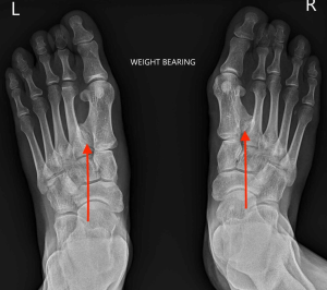
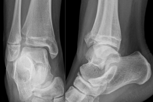
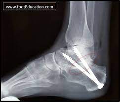
A subtalar joint arthrodesis is a surgical procedure in which the subtalar joint, which is located between the heel bone (calcaneus) and the ankle bone (talus), is fused together to create a single, solid bone. This procedure is typically performed to treat conditions such as severe arthritis, instability, or deformity of the subtalar joint.

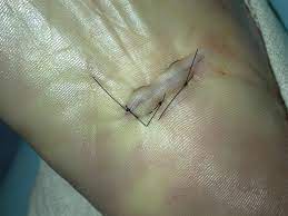
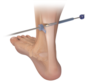
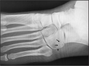
During the procedure, the surgeon makes an incision on the side of the foot and removes the damaged cartilage from the subtalar joint. The bones are then positioned in the desired alignment and held in place with screws or other hardware. Over time, the bones grow together and fuse into a single, solid bone.

Subtalar joint arthrodesis is typically performed under general anesthesia and requires a period of immobilization in a cast or brace. Physical therapy is also recommended to help regain strength and mobility in the affected foot. While this procedure can be highly effective in treating certain conditions, it does limit the range of motion in the foot and ankle and may lead to an increased risk of arthritis in adjacent joints over time. It is important to discuss the potential risks and benefits of subtalar joint arthrodesis with a qualified healthcare professional before undergoing the procedure.

Instep fasciotomy (plantar fasciitis surgery)

An instep plantar fasciotomy is a surgical procedure that involves cutting a portion of the plantar fascia ligament in the foot to relieve tension and pain. The plantar fascia is a thick band of tissue that runs along the bottom of the foot, connecting the heel bone to the toes. When this tissue becomes inflamed or irritated, it can cause a condition called plantar fasciitis, which is characterized by pain and stiffness in the heel and sole of the foot.

During an instep plantar fasciotomy, the surgeon makes a small incision on the top of the foot, near the base of the toes, and cuts a portion of the plantar fascia ligament. This reduces tension on the ligament and relieves pressure on the affected area. The incision is typically closed with stitches, and the foot is immobilized in a cast or boot for several weeks to allow for proper healing.

While instep plantar fasciotomy can be an effective treatment for plantar fasciitis, it is typically reserved for cases that have not responded to non-surgical treatments, such as rest, stretching, and physical therapy. As with any surgical procedure, there are risks and potential complications associated with instep plantar fasciotomy, and it is important to discuss these with a qualified healthcare professional before undergoing the procedure.

Gastrocnemius recession (calf release)

A gastrocnemius recession, also known as gastrocnemius lengthening or calf muscle release, is a surgical procedure in which the gastrocnemius muscle is partially cut to relieve tension and tightness in the calf. This procedure is typically performed to treat a condition called equinus deformity, which is characterized by limited ankle dorsiflexion (the ability to pull the foot upward toward the shin).

During the procedure, a small incision is made in the skin over the gastrocnemius muscle, and the muscle is partially cut or released from its attachment to the heel bone (calcaneus). This allows the muscle to stretch more easily and reduces tension on the Achilles tendon and plantar fascia.

Gastrocnemius recession is often performed in combination with other procedures, such as Achilles tendon lengthening or plantar fascia release, to address the underlying cause of equinus deformity. Recovery from the procedure typically involves a period of immobilization and physical therapy to regain strength and flexibility in the affected leg. As with any surgery, there are risks and potential complications associated with gastrocnemius recession, and it is important to discuss these with a qualified healthcare professional before undergoing the procedure.

Clinodactyly

Clinodactyly is a medical term describing the curvature of a digit (a finger or toe) in the plane of the palm, most commonly the fifth finger (the “little finger”) towards the adjacent fourth finger (the “ring finger”).

It is a fairly common isolated anomaly which often goes unnoticed, but also occurs in combination with other abnormalities in certain genetic syndromes.[1] The term is from the Ancient Greek κλίνειν klínein ‘to bend’ and δάκτυλος dáktulos ‘digit’.

Nutcracker fracture

A nutcracker fracture is a type of foot fracture that occurs when the cuboid bone, located on the outer side of the foot, is crushed between the heel bone and the fourth and fifth metatarsal bones.

This type of fracture can occur as a result of a sudden injury, such as a fall or a direct blow to the foot, or from overuse or repetitive stress on the foot. Symptoms of a nutcracker fracture of the cuboid may include pain, swelling, bruising, and difficulty bearing weight on the affected foot.

Diagnosis is typically made through a physical exam and imaging tests, such as X-rays or CT scans. Treatment for a nutcracker fracture of the cuboid may depend on the severity of the fracture, but generally involves immobilizing the affected foot with a cast or brace and allowing time for the bone to heal. In some cases, surgery may be necessary to realign the fractured bone and stabilize the foot. Physical therapy may also be recommended to help restore range of motion and strength to the affected foot.

Valleix’s sign

Valleix’s sign is another diagnostic test that can be used to assess nerve function in the peripheral nervous system. It involves applying pressure or pinching along the course of a nerve to identify areas of tenderness or pain.

In the case of tarsal tunnel syndrome, the Valleix sign can be used to identify areas of nerve irritation or damage along the course of the posterior tibial nerve. The healthcare provider will apply pressure or pinch along the nerve pathway, starting at the ankle and moving up the leg, while asking the patient if they feel any pain or discomfort.

If the patient experiences pain or tenderness along the course of the posterior tibial nerve, this can be an indication of nerve irritation or damage. However, like Tinel’s sign, the Valleix sign is not always definitive, and additional diagnostic tests may be needed to confirm the diagnosis.

In general, a combination of diagnostic tests and a thorough medical history and physical exam are used to diagnose tarsal tunnel syndrome. Treatment may involve rest, ice, physical therapy, and other conservative measures, and in some cases, surgery may be necessary to relieve pressure on the affected nerve.

Polymetatarsia

Polymetatarsia is a rare condition in which there are more than five metatarsal bones in the forefoot. The most common type of polymetatarsia involves an additional metatarsal bone between the fourth and fifth metatarsals, which is called a “supernumerary metatarsal.” This extra bone can cause pain, discomfort, and difficulty with shoe fitting.

Polymetatarsia may be congenital (present at birth) or acquired (due to injury or disease). It can be diagnosed through physical examination, X-rays, and other imaging studies. Treatment options include wearing custom orthotics or padding to relieve pressure on the foot, modifying shoes to accommodate the extra bone, or surgical removal of the supernumerary metatarsal. The best treatment option depends on the individual’s symptoms and the severity of the condition.

Progressive collapsing flatfoot deformity – Classification system

Progressive collapsing flatfoot deformity can be classified according to the severity of the deformity, as well as the underlying cause. There are several classification systems used by healthcare providers to describe the different stages of the condition. Here are two common classification systems:

  1. Johnson and Strom’s classification system:
  • Stage I: The foot is flat when bearing weight, but the arch returns when the foot is lifted off the ground.
  • Stage II: The arch does not return when the foot is lifted off the ground, but the foot is still flexible.
  • Stage III: The foot is rigid, and there is significant arthritis and deformity.
  1. Myerson’s classification system:
  • Stage I: The hindfoot is in a valgus position (tilted outward) and the arch is slightly flattened.
  • Stage II: The hindfoot is in a valgus position, the arch is more severely flattened, and there is evidence of instability in the ankle joint.
  • Stage III: The hindfoot is in a valgus position, the arch is severely flattened, and there is significant ankle joint arthritis.

The classification system used will depend on the healthcare provider’s preference and the specific needs of the patient.

Tillaux fracture

A Tillaux fracture is a specific type of ankle fracture that occurs in children and adolescents. It is caused by a twisting injury to the ankle and is characterized by a fracture of the lateral aspect of the tibial epiphysis.

The tibial epiphysis is the area of developing bone at the end of the tibia that contributes to the growth of the bone. The lateral aspect of the tibial epiphysis is where the fibula bone attaches to the tibia. When a twisting force is applied to the ankle, it can cause the fibula to pull on the lateral aspect of the tibial epiphysis, resulting in a Tillaux fracture.

Symptoms of a Tillaux fracture may include pain, swelling, and difficulty bearing weight on the affected foot. Treatment typically involves immobilization of the ankle with a cast or brace to allow the fracture to heal. In some cases, surgery may be necessary to realign and stabilize the fractured bone.

If left untreated or improperly treated, a Tillaux fracture can lead to long-term complications such as chronic pain, instability of the ankle joint, and an increased risk of developing arthritis in the ankle. Early and appropriate treatment is important for the best possible outcome.

Pott’s fracture

Pott’s fracture, also known as a bimalleolar ankle fracture, is a type of ankle fracture that involves the tibia and fibula bones. It is typically caused by a twisting injury or direct trauma to the ankle.

In a Pott’s fracture, both the medial malleolus (the bony protrusion on the inner side of the ankle) and the lateral malleolus (the bony protrusion on the outer side of the ankle) are fractured. This can cause significant pain, swelling, and difficulty bearing weight on the affected foot.

Treatment for Pott’s fracture typically involves immobilization of the ankle with a cast or brace to allow the bones to heal. In some cases, surgery may be necessary to realign and stabilize the fractured bones. Rehabilitation and physical therapy may also be necessary to regain strength and mobility in the affected ankle.

If left untreated or improperly treated, a Pott’s fracture can lead to long-term complications such as chronic pain, instability of the ankle joint, and an increased risk of developing arthritis in the ankle.