Category: Musculoskeletal

Tibialis anterior tendinopathy

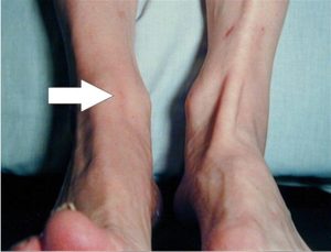
Tibialis anterior tendinopathy is a condition that affects the tibialis anterior tendon, which is located at the front of the ankle and helps to lift the foot upwards. This condition is caused by overuse or repetitive strain on the tendon, and can result in pain, swelling, and weakness in the affected area.

Symptoms of tibialis anterior tendinopathy include pain and tenderness along the front of the ankle, swelling and stiffness in the area, and difficulty lifting the foot upwards. The pain may be worse during physical activity or after prolonged periods of standing or walking.

Treatment for tibialis anterior tendinopathy typically involves rest, ice, compression, and elevation (RICE) to reduce pain and swelling. Non-steroidal anti-inflammatory drugs (NSAIDs) may also be recommended to relieve pain and inflammation. Physical therapy may be recommended to help strengthen the muscles and improve flexibility in the affected area. In severe cases, surgery may be required to repair or replace the damaged tendon.

Prevention of tibialis anterior tendinopathy involves wearing proper footwear and using orthotics or inserts to support the feet and reduce stress on the tendon. Gradually increasing activity levels and avoiding sudden changes in activity levels can also help prevent this condition. It is important to seek medical attention if you experience any symptoms of tibialis anterior tendinopathy, as early diagnosis and treatment can help prevent further damage and improve outcomes.

Tarsal tunnel syndrome

Tarsal tunnel syndrome is a condition in which the tibial nerve, which runs down the leg and into the foot, is compressed as it passes through a narrow tunnel in the ankle called the tarsal tunnel. This can result in pain, tingling, and numbness in the foot and ankle, as well as weakness and muscle atrophy in severe cases.

The symptoms of tarsal tunnel syndrome may be similar to those of other conditions affecting the foot and ankle, such as plantar fasciitis or nerve entrapment in the foot. Therefore, a thorough physical examination, along with imaging tests such as X-rays, MRI, or nerve conduction studies, may be necessary to confirm the diagnosis.

Conservative treatment options for tarsal tunnel syndrome may include rest, icing, physical therapy, and wearing supportive footwear or orthotics. Non-steroidal anti-inflammatory drugs (NSAIDs) may also be prescribed to help relieve pain and inflammation.

In more severe cases or when conservative treatments are not effective, surgery may be necessary to release the pressure on the tibial nerve by removing any structures that may be compressing it.

Early diagnosis and treatment are important for preventing long-term complications such as chronic pain, muscle atrophy, and nerve damage. If you experience persistent foot or ankle pain or notice any unusual symptoms, it is important to seek medical attention to determine the underlying cause and appropriate treatment.

Tarsal coalition

Tarsal coalition is a condition where two or more bones in the foot are abnormally fused together, resulting in limited mobility and pain. It is typically a congenital condition, meaning that it is present at birth, and it often becomes symptomatic during adolescence.

The most common type of tarsal coalition involves the calcaneus bone (heel bone) and the talus bone (bone between the heel bone and the ankle bone), but it can also occur between other bones in the foot.

Symptoms of tarsal coalition may include pain, stiffness, and limited range of motion in the affected foot, particularly during physical activity or standing for prolonged periods. In some cases, the affected foot may also appear to be flat or have a high arch.

Diagnosis of tarsal coalition may involve a physical exam, imaging tests such as X-rays or CT scans, and sometimes, bone scans or MRI.

Treatment for tarsal coalition may depend on the severity of the symptoms and the degree of fusion between the affected bones. Conservative treatment options may include rest, immobilisation with a cast or brace, physical therapy, and non-steroidal anti-inflammatory drugs (NSAIDs) to relieve pain and inflammation.

In more severe cases, surgery may be necessary to remove the abnormal bone or to fuse the affected bones together in a more functional position.

It is important to seek medical attention if you experience persistent foot pain or notice any unusual symptoms, as untreated tarsal coalition can lead to long-term complications such as chronic pain and arthritis.

Talar dome lesion

A talar dome lesion is a type of injury that occurs in the cartilage and bone of the talus, which is the bone in the ankle that connects the foot to the leg. The injury can be caused by a single traumatic event, such as an ankle sprain or fracture, or by repetitive stress on the ankle joint.

The symptoms of a talar dome lesion may include pain, swelling, stiffness, and difficulty bearing weight on the affected ankle. In some cases, the ankle may also feel unstable or give way.

Diagnosis of a talar dome lesion may involve a physical exam, imaging tests such as X-rays, CT scans, or MRI, and in some cases, arthroscopy to visualise the inside of the joint.

Treatment for a talar dome lesion may depend on the severity and location of the injury. Conservative treatments may include rest, ice, compression, and elevation (RICE), immobilisation with a cast or brace, physical therapy, and non-steroidal anti-inflammatory drugs (NSAIDs) to relieve pain and inflammation.

In more severe cases, surgery may be necessary to repair or remove the damaged tissue. This may involve procedures such as micro-fracture, drilling, or cartilage transplantation to stimulate the growth of new cartilage and promote healing.

It is important to seek medical attention if you experience persistent ankle pain or notice any unusual symptoms, as untreated talar dome lesions can lead to long-term complications such as chronic pain, arthritis, and joint instability.

Tailor’s bunion

A tailor’s bunion, also known as a bunionette, is a painful bump that develops on the outside of the foot at the base of the little toe. It is similar to a regular bunion, which develops on the inside of the foot at the base of the big toe.

Tailor’s bunions are usually caused by an inherited structural abnormality in the foot that results in the little toe leaning inward, causing pressure and friction on the outside of the foot. Other factors that can contribute to the development of a tailor’s bunion include wearing tight or poorly fitting shoes, foot injuries, and arthritis.

Symptoms of a tailor’s bunion may include pain, swelling, redness, and a visible bump on the outside of the foot. Treatment for a tailor’s bunion may involve conservative measures such as wearing wider shoes, padding or cushioning the affected area, and taking non-steroidal anti-inflammatory drugs (NSAIDs) to reduce pain and inflammation. In severe cases, surgery may be necessary to correct the underlying structural problem and remove the bony bump.

Prevention of tailor’s bunions involves wearing comfortable, properly fitting shoes that provide enough room for the toes to move freely. Avoiding high heels and shoes with narrow toe boxes can also help reduce the risk of developing a tailor’s bunion. If you experience persistent foot pain or notice any unusual bumps or growths on your feet, it is important to seek medical attention to determine the underlying cause and receive appropriate treatment.

Stress fractures

A stress fracture in the foot is a small crack or break in one of the bones of the foot, typically caused by repetitive stress or overuse. This type of injury is common among athletes, particularly those who participate in high-impact sports such as running, jumping, or dancing.

Some common symptoms of a stress fracture in the foot may include:

  • Pain that worsens with activity and improves with rest
  • Swelling or tenderness in the affected area
  • Bruising or discolouration around the affected area
  • Difficulty bearing weight on the affected foot

If you suspect that you have a stress fracture in your foot, it is important to seek medical attention right away. Treatment may include rest, immobilisation with a cast or brace, and the use of crutches to avoid putting weight on the affected foot. Non-steroidal anti-inflammatory drugs (NSAIDs) may also be recommended to help reduce pain and inflammation.

Rehabilitation exercises and physical therapy may also be recommended to help strengthen the muscles and improve range of motion in the foot. It is important to follow your healthcare provider’s instructions and allow adequate time for your foot to heal, as returning to activity too soon can cause the stress fracture to worsen or lead to additional injuries.

Shin splints

Shin splints, also known as medial tibial stress syndrome, is a common injury that occurs when the muscles and tendons surrounding the shinbone become inflamed. It is often caused by overuse or repetitive stress, such as running or jumping on hard surfaces.

Symptoms of shin splints include pain, tenderness, and swelling along the inner edge of the shinbone. The pain may be mild at first but can become more severe with continued activity.

To prevent shin splints, it is important to gradually increase the intensity and duration of your activities, wear proper footwear that provides adequate cushioning and support, and avoid running or jumping on hard surfaces. Additionally, stretching and strengthening exercises for the lower leg muscles can help prevent shin splints.

If you do develop shin splints, the best treatment is rest. Avoid the activities that caused the injury and allow your legs time to heal. Ice, compression, and elevation (RICE) can also help reduce pain and swelling. Over-the-counter pain medications, such as ibuprofen, can also be used to manage pain.

Once the pain and swelling have subsided, it is important to gradually return to your activities to avoid re-injuring the area. Wearing proper footwear and using orthotics or inserts can also help prevent future episodes of shin splints. If the pain persists despite these measures, it is important to see a healthcare professional for further evaluation and treatment.

Sesamoid injuries

The sesamoid bones are small bones located under the big toe joint in the foot. They play an important role in weight-bearing and movement of the foot. Sesamoid injuries are a common condition that occurs when the sesamoid bones become injured or inflamed.

There are two types of sesamoid injuries:

  1. Sesamoiditis: This is a condition in which the sesamoid bones become inflamed due to overuse or repetitive stress. Symptoms of sesamoiditis include pain, swelling, and tenderness in the affected area.
  2. Sesamoid Fracture: This is a type of injury that occurs when one or both of the sesamoid bones in the foot are fractured. Symptoms of a sesamoid fracture include severe pain, swelling, and difficulty walking.

To prevent sesamoid injuries, it is important to wear proper footwear that provides adequate cushioning and support, especially if you engage in activities that involve repetitive stress on the foot. In addition, avoid activities that put excessive pressure on the foot, such as jumping and running on hard surfaces.

If you suspect you have a sesamoid injury, it is important to seek medical attention. Treatment for sesamoid injuries may include rest, ice, compression, and elevation (RICE), over-the-counter pain medications, and physical therapy. In severe cases, surgery may be necessary.

Posterior tibial tendon dysfunction

Posterior tibial tendon dysfunction (PTTD) is a condition that occurs when the posterior tibial tendon, which runs along the inside of the ankle and foot, becomes damaged or inflamed. The posterior tibial tendon is responsible for supporting the arch of the foot and helping to maintain proper alignment and stability.

PTTD is often caused by overuse or repetitive stress on the posterior tibial tendon, which can lead to small tears or degeneration of the tendon. Other factors that may contribute to PTTD include obesity, flat feet, high-impact activities, and certain medical conditions such as rheumatoid arthritis.

Symptoms of PTTD may include pain and swelling along the inside of the ankle and foot, particularly during and after physical activity, as well as a gradual flattening of the arch of the foot and a shifting of the heel bone outward.

Treatment for PTTD typically involves a combination of non-surgical interventions such as rest, ice, compression, elevation, and physical therapy to help reduce pain and inflammation and improve strength and flexibility in the affected foot and ankle. Orthotic devices such as arch supports or braces may also be recommended to help support the foot and relieve stress on the posterior tibial tendon.

In more severe cases of PTTD, surgical intervention may be necessary to repair or reconstruct the damaged tendon, restore proper alignment of the foot, and improve overall foot and ankle function. It is important to consult with a healthcare professional to determine the most appropriate treatment plan for your specific condition and individual health needs.

Plantar plate tear

The plantar plate is a fibrocartilaginous structure located on the bottom of the foot, which serves to support and stabilize the metatarsophalangeal (MTPJ) joints. A plantar plate tear is a common injury that occurs when the ligamentous fibres of the plantar plate are partially or completely torn.

Plantar plate tears are typically caused by repetitive stress or trauma to the foot, such as from running, jumping, or other high-impact activities. They may also occur as a result of degenerative changes associated with aging or certain medical conditions, such as rheumatoid arthritis.

Symptoms of a plantar plate tear can include pain, swelling, and tenderness in the ball of the foot, particularly when weight-bearing or walking. There may also be a feeling of instability or a “popping” sensation in the affected joint.

Physiotherapy treatment for plantar plate tears may include rest, ice, and immobilisation to reduce pain and inflammation. Manual therapy techniques, such as massage and joint mobilisation, may also be used to improve range of motion and reduce stiffness in the affected area.

Strengthening and stretching exercises can help improve foot and ankle strength and flexibility, and may include exercises to target the plantar fascia and other supporting structures of the foot.

In some cases, custom orthotics or supportive footwear may also be recommended to help relieve pressure on the affected area and prevent further injury.

Surgical intervention may be necessary in cases of severe or chronic plantar plate tears, particularly if conservative treatment is not effective. It is important to consult with a healthcare professional if you suspect you have a plantar plate tear, as early diagnosis and treatment can help prevent further damage and improve overall outcomes.