Category: Musculoskeletal

Coughlin & Shurnas classification system for hallux rigidus


The Coughlin and Shurnas classification system is used to categorize different stages of hallux rigidus, a degenerative condition of the big toe joint (first metatarsophalangeal joint) that leads to stiffness and limited motion. This classification system helps surgeons and medical professionals assess the severity of hallux rigidus and determine appropriate treatment strategies.

The Coughlin and Shurnas classification for hallux rigidus consists of four stages:

Stage 0: No arthritic changes are present. The joint has normal or near-normal function, and there is no pain or stiffness.

Stage 1: Mild or early-stage hallux rigidus. Some joint space narrowing and osteophyte formation (bone spurs) are present. There might be mild pain and stiffness with movement.

Stage 2: Moderate hallux rigidus. The joint space is further reduced, and larger osteophytes are present. Joint motion is more restricted, and pain is more pronounced, especially during push-off while walking.

Stage 3: Severe hallux rigidus. The joint space is significantly narrowed or obliterated, and osteophytes are prominent. Joint motion is severely limited, and the patient experiences significant pain and dysfunction, even during walking.

Hardcastle & Myerson classification for Lisfranc injuries

The Hardcastle and Myerson classification system is used to categorize injuries to the Lisfranc joint complex, which involves the tarsometatarsal joints in the midfoot. This classification system helps surgeons and medical professionals assess the severity of Lisfranc injuries and guide treatment decisions. Lisfranc injuries can range from mild sprains to severe fractures and dislocations.

The Hardcastle and Myerson classification system is divided into three main categories, which are further sub-divided based on the severity of the injury:

Type A: Homolateral Fracture Dislocation

  1. Type A1: Fracture of the base of the second metatarsal.
  2. Type A2: Fracture of the first and second metatarsals.
  3. Type A3: Fracture of all three medial metatarsals.

Type B: Isolated Lisfranc Ligament Injuries

  1. Type B1: Sprain or rupture of the Lisfranc ligament with or without fractures.

Type C: Divergent Dislocation

  1. Type C1: Dislocation of one or two metatarsals.
  2. Type C2: Dislocation of three or more metatarsals.

Each type is associated with varying degrees of instability and severity. Treatment options and outcomes depend on the specific type of Lisfranc injury.

It’s important to note that Lisfranc injuries can be complex and may require careful evaluation by a medical professional, often including imaging studies like X-rays or CT scans. Treatment can range from non-surgical approaches (such as casting or bracing) for less severe injuries to surgical intervention (such as stabilization with screws or plates) for more severe injuries.

If you suspect a Lisfranc injury, it’s crucial to seek prompt medical attention to ensure an accurate diagnosis and appropriate management to prevent potential long-term complications, such as chronic pain and instability in the midfoot.

Ankle sprain classification

Ankle sprains are common injuries that occur when the ligaments around the ankle joint are stretched or torn due to excessive force or twisting. There are various classification systems used to describe and categorize ankle sprains based on the severity and location of the injury. One of the commonly used classification systems is the Grading System:

  1. Grade I (Mild): In a Grade I ankle sprain, the ligaments are stretched but not torn. There may be mild pain, swelling, and minimal loss of function. The ankle joint remains stable.
  2. Grade II (Moderate): A Grade II ankle sprain involves partial tearing of the ligaments. This can cause moderate pain, swelling, and difficulty walking. The ankle joint may feel unstable.
  3. Grade III (Severe): In a Grade III ankle sprain, the ligaments are completely torn. This results in significant pain, swelling, bruising, and instability of the ankle joint. Walking and weight-bearing are usually impaired.

Another classification system used specifically for lateral ankle sprains is the Anatomic Grading System, which focuses on the specific ligaments involved:

  1. Stage I: Mild sprain with microscopic tearing of the ligament fibers.
  2. Stage II: Moderate sprain with partial tearing of the ligament fibers.
  3. Stage III: Severe sprain with complete rupture of the ligament.

Additionally, there’s a classification system that considers the location of the injury within the lateral ankle ligament complex:

  1. Anterior Talofibular Ligament (ATFL) Sprain: The most common type of ankle sprain, often occurring in inversion injuries. It involves the ligament on the front and outside of the ankle.
  2. Calcaneofibular Ligament (CFL) Sprain: This involves the ligament on the outside of the ankle and is commonly associated with more severe sprains.
  3. Posterior Talofibular Ligament (PTFL) Sprain: This ligament, located on the back of the ankle, is less frequently injured.

Lauge-Hansen classification system for ankle fractures

The Lauge-Hansen classification system is a widely used method for describing and categorizing different types of ankle fractures based on the mechanism of injury. It was developed by Danish orthopaedic surgeon Lauge-Hansen in the mid-20th century.

This classification system takes into account two main factors: the position of the foot at the time of injury (pronation or supination) and the direction of the force applied (medial or lateral).

The Lauge-Hansen classification system consists of four main fracture patterns:

  1. Supination-Adduction (SA) injuries: These occur when the foot is in a supinated (turned inward) position and a force is applied in an adduction (inward) direction. This typically results in a fracture of the lateral malleolus (fibula) and possible rupture of the deltoid ligament on the medial side of the ankle.
  2. Supination-External Rotation (SER) injuries: These occur when the foot is in a supinated position and a force is applied in an external rotation (outward) direction. This can result in a fracture of the fibula, followed by a rupture of the anterior talofibular ligament and the calcaneofibular ligament.
  3. Pronation-Abduction (PA) injuries: These occur when the foot is in a pronated (turned outward) position and a force is applied in an abduction (outward) direction. This can lead to a fracture of the medial malleolus (tibia) and possible rupture of the lateral ligaments.
  4. Pronation-External Rotation (PER) injuries: These occur when the foot is in a pronated position and a force is applied in an external rotation direction. This can result in a fracture of the medial malleolus, followed by rupture of the deltoid ligament and the syndesmosis (the ligaments that hold the tibia and fibula together).

The Lauge-Hansen classification system helps surgeons and medical professionals better understand the specific patterns of ankle fractures, which can guide treatment decisions and surgical approaches.

Dorsal osseous compression syndrome (DOCS)

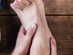
Dorsal osseous compression syndrome (DOCS), also known as dorsal compression syndrome or dorsalis pedis compression syndrome, is a medical condition characterized by compression of the dorsal (top) surface of the foot, usually by adjacent bones or structures. It can cause pain, discomfort, and other symptoms in the affected foot.

The condition typically occurs due to abnormal pressure or compression on the dorsum of the foot, which can result from various causes, including:

  1. Tight footwear: Wearing shoes that are too tight, narrow, or constrictive can compress the bones and soft tissues on the top of the foot, leading to dorsal osseous compression syndrome.
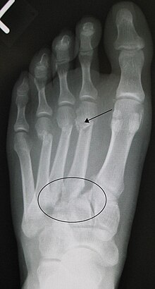
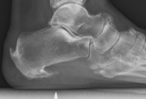
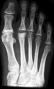
  2. Bone deformities: Some individuals may have anatomical variations in their foot bones, such as prominent dorsal bones or accessory bones, which can lead to compression of nearby structures.
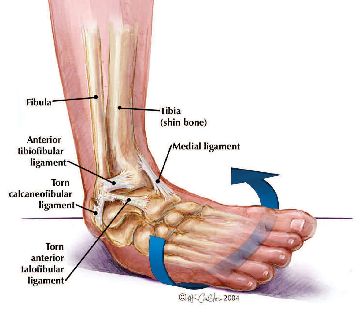
  3. Trauma: Previous injuries or trauma to the foot, such as fractures or dislocations, can result in changes in the alignment of the foot bones, leading to dorsal compression syndrome.
  4. Overuse or repetitive activities: Repeated activities that involve excessive dorsiflexion of the foot, such as running, jumping, or wearing high-heeled shoes for prolonged periods, can cause compression of the dorsum of the foot.

Symptoms of foot dorsal osseous compression syndrome may include pain, tenderness, swelling, bruising, and limited range of motion in the affected foot. The pain may worsen with activity, walking, or wearing tight shoes, and may improve with rest and elevation of the foot.

Treatment options for foot dorsal osseous compression syndrome depend on the underlying cause and severity of the condition. Conservative treatments may include rest, ice, elevation, compression, wearing properly fitting footwear, and avoiding activities that exacerbate the symptoms. Orthotic devices, such as padding or shoe inserts, may also be recommended to alleviate pressure on the affected area.

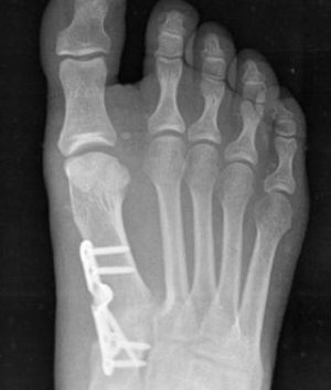
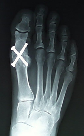
In some cases, if conservative treatments are ineffective, more advanced interventions may be required, such as corticosteroid injections, physical therapy, or in rare cases, surgical interventions to address any bone deformities or structural abnormalities causing the compression.

If you suspect you may have foot dorsal osseous compression syndrome or are experiencing foot pain or discomfort, it is important to consult with a healthcare professional for a proper diagnosis and appropriate treatment plan tailored to your individual needs.

Sinus tarsi syndrome

Sinus tarsi syndrome is a condition that affects the foot and ankle. It is characterized by pain and discomfort in the sinus tarsi, which is a small bony canal located between the talus bone (ankle bone) and the calcaneus bone (heel bone). The sinus tarsi serves as a passage for nerves, blood vessels, and ligaments that are important for foot and ankle function.

Sinus tarsi syndrome is typically caused by injury or trauma to the foot and ankle, such as ankle sprains, repetitive overuse, or chronic instability. Other potential causes can include arthritis, ligamentous laxity, or anatomical variations that lead to compression or irritation of the nerves or tissues within the sinus tarsi.

Symptoms of sinus tarsi syndrome may include:

  1. Pain: Pain in the lateral (outer) aspect of the foot, specifically in the area of the sinus tarsi, is the hallmark symptom of this condition. The pain may be sharp or dull and may worsen with weight-bearing activities or prolonged standing.
  2. Swelling: Swelling around the sinus tarsi may be present, although it is usually mild compared to other foot and ankle conditions.
  3. Tenderness: Tenderness to touch over the sinus tarsi area may be present, and pressure on the area may exacerbate the pain.
  4. Instability: Some patients with sinus tarsi syndrome may experience a feeling of instability or a sense of “giving way” in the foot or ankle.
  5. Limited range of motion: Reduced range of motion in the ankle joint may be observed, particularly with movements that involve inversion (inward rolling) or eversion (outward rolling) of the foot.

Diagnosis of sinus tarsi syndrome typically involves a thorough clinical evaluation by a healthcare provider, including a physical examination, assessment of medical history, and imaging studies such as X-rays or MRI to rule out other potential causes of foot and ankle pain.

Treatment for sinus tarsi syndrome may include conservative measures such as rest, ice, compression, and elevation (RICE), nonsteroidal anti-inflammatory drugs (NSAIDs), orthotics or shoe modifications, physical therapy, and activity modification. In some cases, corticosteroid injections may be used to reduce inflammation and alleviate pain. If conservative measures are not effective, more advanced treatments such as extracorporeal shockwave therapy, prolotherapy, or platelet-rich plasma (PRP) injections may be considered. In rare cases, surgical intervention may be necessary to address any underlying structural issues or persistent symptoms.

It is important to consult with a qualified healthcare provider for an accurate diagnosis and appropriate treatment plan if you suspect you may have sinus tarsi syndrome or are experiencing foot and ankle pain.

Forestier’s disease

Forestier’s disease can affect various parts of the body, including the spine, pelvis, hip, and knee, but it is less common for it to affect the foot. However, in rare cases, Forestier’s disease can cause abnormal bone growth in the foot, leading to stiffness, pain, and difficulty with movement.

The symptoms of Forestier’s disease in the foot may include:

  • Stiffness and limited range of motion in the affected joint(s)
  • Pain that worsens with activity
  • Swelling or tenderness around the affected area
  • Deformity or abnormal bone growth in the foot

Treatment for Forestier’s disease in the foot may include nonsteroidal anti-inflammatory drugs (NSAIDs) for pain and inflammation, physical therapy to improve range of motion and mobility, and in severe cases, surgery to remove or reshape the abnormal bone growth. It’s important to consult with a healthcare provider for proper diagnosis and treatment of Forestier’s disease in the foot.

Lapidus procedure

The Lapidus procedure is a surgical procedure used to treat hallux valgus, a condition in which the big toe drifts away from the midline of the foot, causing a bunion. It involves fusing the first metatarsal bone to the medial cuneiform bone in the midfoot to correct the alignment of the bones and reduce the deformity. This procedure is typically reserved for cases of severe hallux valgus or for patients who have not responded to more conservative treatments.

During the procedure, the surgeon makes an incision on the top of the foot and removes a small piece of bone from the base of the first metatarsal. The metatarsal bone is then repositioned and fixed in place with screws or a plate to hold it in the desired alignment. Over time, the bones grow together and form a solid fusion, which helps to stabilize the midfoot and reduce the severity of the bunion.

After the surgery, the patient may need to wear a cast or brace for several weeks to protect the foot and allow the bones to fuse together. Physical therapy may also be recommended to help improve strength, flexibility, and range of motion in the affected foot. While the Lapidus procedure can be highly effective in correcting hallux valgus, it does require a period of immobilization and recovery, and may have some potential risks and complications, such as non-union, nerve injury, or infection. It is important to discuss the potential risks and benefits of the Lapidus procedure with a qualified healthcare professional before undergoing the procedure.

Freiberg’s infarction

Freiberg’s infarction, also known as Freiberg’s disease or osteochondrosis of the metatarsal head, is a condition in which there is a loss of blood supply (avascular necrosis) to the head of the metatarsal bone in the foot. This can cause pain, swelling, and stiffness in the affected foot, and may lead to arthritis or joint deformity over time.

The exact cause of Freiberg’s infarction is not fully understood, but it is believed to be related to repeated trauma or injury to the metatarsal head, which can disrupt the blood supply to the bone. The condition is most commonly seen in adolescent girls and young women, and may be associated with wearing high-heeled shoes or engaging in activities that involve repetitive impact on the foot, such as running or jumping.

Treatment for Freiberg’s infarction typically involves rest, ice, and anti-inflammatory medication to reduce pain and inflammation in the affected foot. Immobilization in a cast or boot may also be recommended to allow the bone to heal. In more severe cases, surgery may be necessary to remove damaged bone or reshape the joint to reduce pain and improve function.

Overall, the prognosis for Freiberg’s infarction depends on the severity of the condition and the effectiveness of the chosen treatment. With appropriate care, many patients are able to manage their symptoms and maintain good foot function over time.

First metatarsophalangeal joint arthrodesis

A first metatarsophalangeal joint (1st MTPJ) arthrodesis is a surgical procedure that involves fusing the bones of the big toe (metatarsal) and the first bone of the toe (phalanx) together to create a solid joint. This procedure is typically performed to treat advanced arthritis, instability, or deformity of the 1st MTPJ.

During the procedure, the surgeon makes an incision on the top of the foot and removes the damaged joint surfaces of the metatarsal and phalanx bones. The bones are then placed in the desired alignment and held in place with screws, pins, or plates until they fuse together. Over time, the bones grow together and form a solid joint, which can help relieve pain and improve the stability and function of the foot.

After the surgery, the foot is immobilized in a cast or boot for several weeks, and physical therapy may be recommended to help regain strength and mobility in the affected foot. While 1st MTPJ arthrodesis can be highly effective in treating certain conditions, it does limit the range of motion in the big toe and may affect the gait or balance of the patient. It is important to discuss the potential risks and benefits of 1st MTPJ arthrodesis with a qualified healthcare professional before undergoing the procedure.