Category: Musculoskeletal

Köhler disease

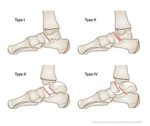
Köhler disease, also known as osteochondrosis of the navicular bone, is a rare condition that affects the navicular bone in the foot. It is most commonly seen in children between the ages of 5 and 10 years old, and is more common in boys than girls.

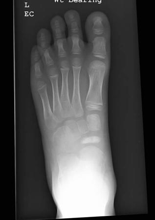
The exact cause of Köhler disease is unknown, but it is believed to be related to a disruption in blood flow to the navicular bone. This can lead to a decrease in bone density and the development of small fractures in the bone.

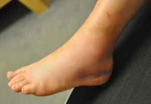
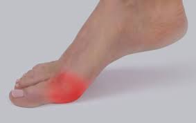
Symptoms of Köhler disease typically include pain and swelling in the midfoot, particularly on the top of the foot. The affected foot may also appear flattened or widened, and there may be a limp or difficulty walking.

Treatment for Köhler disease usually involves rest and immobilization of the foot with a cast or brace to allow the bone to heal. Pain relievers may also be prescribed to manage discomfort. In rare cases, surgery may be necessary to remove damaged tissue or realign the bones in the foot. With appropriate treatment, most children with Köhler disease recover fully and have no long-term complications.

Anterior talofibular ligament (ATFL) rupture

An anterior talofibular ligament (ATFL) rupture can be classified according to the severity of the injury, as follows:

  1. Grade I: Mild sprain with minimal or no ligament fiber tear. This type of injury usually causes mild swelling and pain, but the patient can still walk normally.
  2. Grade II: Moderate sprain with partial ligament fiber tear. This type of injury causes moderate swelling, pain, and instability of the ankle joint. The patient may experience difficulty walking and may need crutches or a brace to support the ankle.
  3. Grade III: Severe sprain with complete ligament tear. This type of injury causes severe swelling, pain, and instability of the ankle joint. The patient is usually unable to bear weight on the affected ankle and may require surgery to repair the torn ligament.

It is important to note that the severity of the injury does not always correlate with the level of pain or disability experienced by the patient. In some cases, even a mild sprain can cause significant pain and disability, while in other cases, a severe sprain may cause little pain or disability.

Weber classification for fibular fractures

Weber fractures are a classification system for fractures of the ankle, specifically the fibula bone. The Weber classification system is based on the location of the fracture relative to the ankle joint and the degree of displacement of the fracture.

There are three types of Weber fractures:

  • Weber A: This is a fracture of the fibula that occurs below the level of the ankle joint. The ankle joint itself is not affected. The fracture may be non-displaced (the bone is still aligned properly) or displaced (the bone is out of alignment). This type of fracture is usually treated with immobilization and rest.
  • Weber B: This is a fracture of the fibula that occurs at the level of the ankle joint. The ankle joint is also affected, as the fracture extends into the ligaments that connect the fibula to the tibia bone. This type of fracture is typically treated with immobilization, rest, and sometimes surgery to realign the bones and stabilize the joint.
  • Weber C: This is a fracture of the fibula that occurs above the level of the ankle joint, often at the level of the syndesmosis (the joint between the tibia and fibula bones). The ankle joint is not usually affected, but there may be significant ligament damage. This type of fracture is often treated with surgery to realign the bones and stabilize the joint.

Overall, the Weber classification system is a useful tool for healthcare professionals in assessing and managing ankle fractures. Treatment options for Weber fractures may include immobilization with a cast or brace, surgery to realign the bones and stabilize the joint, and physical therapy to restore range of motion and strength to the ankle.

Salter-Harris classification for growth plate fractures

The Salter-Harris classification is a system used to classify fractures that involve the growth plate, also known as the epiphyseal plate, in pediatric patients. The growth plate is a cartilage-rich area at the ends of long bones that allows for bone growth and development.

The Salter-Harris classification divides growth plate fractures into five categories, based on the location and extent of the fracture:

Type I: This is a transverse fracture that runs through the growth plate, separating the epiphysis (the end of the bone) from the metaphysis (the shaft of the bone). This is the most common type of growth plate fracture and is usually treated with immobilization and close monitoring.

Type II: This is an oblique fracture that runs through the growth plate and into the metaphysis. This type of fracture is also treated with immobilization and monitoring, and may require more frequent follow-up to ensure proper healing.

Type III: This is a fracture that runs through the growth plate and into the epiphysis. This type of fracture may require more aggressive treatment, such as surgery, to prevent long-term complications such as growth disturbances or joint deformities.

Type IV: This is a fracture that runs through the growth plate, the epiphysis, and the metaphysis. This type of fracture is relatively rare and may require surgical intervention to prevent long-term complications.

Type V: This is a crush injury to the growth plate that results in damage to the cells responsible for bone growth. This type of fracture is also relatively rare and may require surgical intervention to prevent growth disturbances.

The Salter-Harris classification is a useful tool for healthcare professionals in assessing and managing growth plate fractures in pediatric patients. Treatment options for growth plate fractures may include immobilization, closed reduction (manipulation of the bones to restore proper alignment), and surgery in some cases.

Overall, prompt and appropriate treatment of growth plate fractures is important to minimize the risk of long-term complications and ensure proper bone growth and development.

Sanders classification for calcaneal fractures

The Sanders classification is a system used to categorize calcaneal fractures, which are fractures of the heel bone in the foot. The classification was developed by Dr. Roy W. Sanders, an American orthopedic surgeon, in 1993.

The Sanders classification divides calcaneal fractures into four categories, based on the location and severity of the fracture:

Type I: This is a simple, non-displaced fracture of the posterior calcaneal tuberosity, which is a bony protrusion at the back of the heel bone. This type of fracture is considered to be relatively minor, and is often treated non-surgically with immobilization and rest.

Type II: This is a displaced fracture of the posterior calcaneal facet, which is the portion of the heel bone that articulates with the talus bone in the ankle joint. This type of fracture can result in joint incongruity (misalignment) and can be associated with long-term complications.

Type III: This is a fracture involving both the posterior calcaneal facet and the middle facet of the heel bone. This type of fracture is more severe than Type II, and is associated with a higher risk of complications.

Type IV: This is a fracture involving the entire calcaneus bone, and is the most severe type of calcaneal fracture. This type of fracture can result in significant joint incongruity and is associated with a high risk of long-term complications.

The Sanders classification can be useful in guiding treatment decisions for calcaneal fractures, as the severity and location of the fracture can impact the likelihood of complications such as joint incongruity and post-traumatic arthritis. Treatment options for calcaneal fractures may include immobilization with a cast or brace, surgery to realign the bones and stabilize the joint, and in some cases, joint replacement surgery.

Overall, the Sanders classification is a valuable tool for healthcare professionals in assessing and managing calcaneal fractures, and can help improve patient outcomes through more targeted and effective treatment.

Hawkins classification for talar fractures

The Hawkins classification is a system used to categorize talar fractures, which are fractures of the talus bone in the ankle. The classification was developed by Dr. Herbert Hawkins, an American orthopedic surgeon, in 1970.

The Hawkins classification divides talar fractures into four categories, based on the location and severity of the fracture:

Type I: This is a non-displaced fracture of the talar neck, which is the narrow portion of the talus bone between the body of the talus and the ankle joint. The blood supply to the talus is usually preserved in this type of fracture.

Type II: This is a displaced fracture of the talar neck, which can result in damage to the blood supply to the talus. This type of fracture is considered to be more severe than Type I.

Type III: This is a fracture of the body of the talus, which is the large, rounded portion of the bone that forms the ankle joint. This type of fracture is often associated with significant damage to the blood supply to the talus, and can result in avascular necrosis (death of bone tissue due to loss of blood supply) of the talus.

Type IV: This is a fracture of the posterior process of the talus, which is a small projection on the back of the talus bone. This type of fracture is less common than the other three types, and is generally considered to be less severe.

The Hawkins classification can be useful in guiding treatment decisions for talar fractures, as the severity and location of the fracture can impact the likelihood of complications such as avascular necrosis. Treatment options for talar fractures may include immobilization with a cast or brace, surgery to realign the bones and stabilize the joint, and in some cases, joint replacement surgery.

Overall, the Hawkins classification is a valuable tool for healthcare professionals in assessing and managing talar fractures, and can help improve patient outcomes through more targeted and effective treatment.

Why won’t my ankle sprain heal?

An ankle sprain can take anywhere from a few days to several weeks to heal, depending on the severity of the sprain. However, there are a few reasons why an ankle sprain may not be healing as quickly as expected, including:

  1. Insufficient Rest: One of the most common reasons for an ankle sprain not healing is insufficient rest. Continuing to put weight on the injured ankle can cause further damage and delay the healing process.
  2. Inadequate Rehabilitation: It’s important to properly rehabilitate the ankle after a sprain, including doing exercises to improve strength, balance, and range of motion. Failure to properly rehabilitate the ankle can result in weakness and instability, making it more susceptible to re-injury.
  3. Repeated Injury: Repeated injury to the same ankle can cause chronic pain and instability, making it difficult for the ankle to heal properly.
  4. Incorrect Diagnosis: In some cases, the initial diagnosis of an ankle sprain may be incorrect. If the ankle does not seem to be healing as expected, it may be necessary to reevaluate the diagnosis and consider other possible underlying conditions.
  5. Underlying Conditions: Some underlying medical conditions, such as arthritis or gout, can make it more difficult for an ankle sprain to heal.

If you are experiencing an ankle sprain that is not healing as quickly as expected, it’s important to see a healthcare provider or a podiatrist to determine the underlying cause of the problem. They can help diagnose the condition and recommend appropriate treatments, such as rest, rehabilitation exercises, or in some cases, surgery, to help improve the healing process and prevent further injury.

Why does my heel hurt?

Heel pain is a common complaint that can be caused by a number of different conditions. Here are some of the most common causes of heel pain:

  1. Plantar Fasciitis: This is a condition where the plantar fascia, a band of tissue that runs along the bottom of the foot, becomes inflamed. This can cause pain in the heel or arch of the foot, especially when taking the first steps in the morning or after periods of inactivity.
  2. Heel Spurs: A heel spur is a bony growth that forms on the heel bone, often as a result of long-term plantar fasciitis. The spur itself is not painful, but it can cause pain by irritating the surrounding tissues.
  3. Achilles Tendinitis: This is a condition where the Achilles tendon, which connects the calf muscles to the heel bone, becomes inflamed. This can cause pain and stiffness in the heel or back of the ankle.
  4. Bursitis: Bursitis is a condition where the bursae, small fluid-filled sacs that cushion the joints, become inflamed. This can cause pain in the heel or other parts of the foot.
  5. Stress Fracture: A stress fracture is a small crack in the bone that can be caused by overuse or repetitive impact. This can cause pain in the heel or other parts of the foot.
  6. Nerve Impingement: A nerve in the foot can become pinched or compressed, leading to pain in the heel or other parts of the foot.

If you are experiencing heel pain, it’s important to see a healthcare provider or a podiatrist to determine the underlying cause of the problem. They can help diagnose the condition and recommend appropriate treatments, such as stretching exercises, orthotics, or physical therapy, to help relieve the pain and improve your mobility.

Webbed toes

Webbed toes, also known as syndactyly, is a condition where two or more toes are fused together by a web of skin and tissue. This can occur in either the feet or the hands, and it is a relatively common condition that affects about 1 in 2,000 to 1 in 3,000 births.

In most cases, webbed toes are not harmful and do not cause any problems with walking or other activities. However, in some cases, the condition may cause difficulty wearing shoes or may be associated with other health problems.

Webbed toes can be treated through surgery, which involves separating the fused digits and reconstructing the skin and soft tissue to create separate toes. The timing of the surgery may depend on the severity of the webbing and whether it is affecting the child’s ability to walk or perform other activities.

It’s important to note that in some cases, webbed toes may be a sign of an underlying genetic condition or syndrome, so it’s important to consult with a healthcare provider if you or your child has webbed toes. They can evaluate the condition and determine whether any further testing or treatment is necessary.

Turf toe

Turf toe is a common injury that occurs when the big toe is forcibly bent upward, causing damage to the ligaments and soft tissues around the joint. It is most commonly seen in athletes who play sports on artificial turf, hence the name “turf toe.”

Symptoms of turf toe include pain, swelling, and stiffness in the affected toe, as well as difficulty walking or bearing weight on the foot. In severe cases, the joint may be dislocated or fractured.

Treatment for turf toe typically involves rest, ice, compression, and elevation (RICE) to reduce pain and swelling. Anti-inflammatory medications may also be recommended to help manage symptoms. In some cases, a stiff-soled shoe or special orthotic device may be prescribed to limit movement of the affected joint and prevent further injury.

Physical therapy may also be recommended to help restore range of motion and strength to the affected foot and prevent future injuries. In rare cases where the joint is severely damaged, surgery may be necessary to repair or reconstruct the ligaments and soft tissues around the joint.