Category: Fractures

Weber classification for fibular fractures

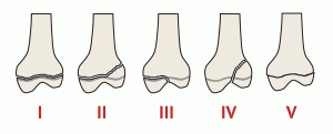
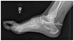
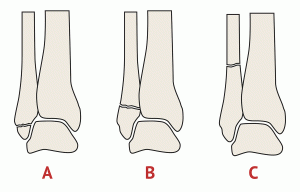
Weber fractures are a classification system for fractures of the ankle, specifically the fibula bone. The Weber classification system is based on the location of the fracture relative to the ankle joint and the degree of displacement of the fracture.

There are three types of Weber fractures:

  • Weber A: This is a fracture of the fibula that occurs below the level of the ankle joint. The ankle joint itself is not affected. The fracture may be non-displaced (the bone is still aligned properly) or displaced (the bone is out of alignment). This type of fracture is usually treated with immobilization and rest.
  • Weber B: This is a fracture of the fibula that occurs at the level of the ankle joint. The ankle joint is also affected, as the fracture extends into the ligaments that connect the fibula to the tibia bone. This type of fracture is typically treated with immobilization, rest, and sometimes surgery to realign the bones and stabilize the joint.
  • Weber C: This is a fracture of the fibula that occurs above the level of the ankle joint, often at the level of the syndesmosis (the joint between the tibia and fibula bones). The ankle joint is not usually affected, but there may be significant ligament damage. This type of fracture is often treated with surgery to realign the bones and stabilize the joint.

Overall, the Weber classification system is a useful tool for healthcare professionals in assessing and managing ankle fractures. Treatment options for Weber fractures may include immobilization with a cast or brace, surgery to realign the bones and stabilize the joint, and physical therapy to restore range of motion and strength to the ankle.

Salter-Harris classification for growth plate fractures

The Salter-Harris classification is a system used to classify fractures that involve the growth plate, also known as the epiphyseal plate, in pediatric patients. The growth plate is a cartilage-rich area at the ends of long bones that allows for bone growth and development.

The Salter-Harris classification divides growth plate fractures into five categories, based on the location and extent of the fracture:

Type I: This is a transverse fracture that runs through the growth plate, separating the epiphysis (the end of the bone) from the metaphysis (the shaft of the bone). This is the most common type of growth plate fracture and is usually treated with immobilization and close monitoring.

Type II: This is an oblique fracture that runs through the growth plate and into the metaphysis. This type of fracture is also treated with immobilization and monitoring, and may require more frequent follow-up to ensure proper healing.

Type III: This is a fracture that runs through the growth plate and into the epiphysis. This type of fracture may require more aggressive treatment, such as surgery, to prevent long-term complications such as growth disturbances or joint deformities.

Type IV: This is a fracture that runs through the growth plate, the epiphysis, and the metaphysis. This type of fracture is relatively rare and may require surgical intervention to prevent long-term complications.

Type V: This is a crush injury to the growth plate that results in damage to the cells responsible for bone growth. This type of fracture is also relatively rare and may require surgical intervention to prevent growth disturbances.

The Salter-Harris classification is a useful tool for healthcare professionals in assessing and managing growth plate fractures in pediatric patients. Treatment options for growth plate fractures may include immobilization, closed reduction (manipulation of the bones to restore proper alignment), and surgery in some cases.

Overall, prompt and appropriate treatment of growth plate fractures is important to minimize the risk of long-term complications and ensure proper bone growth and development.

Sanders classification for calcaneal fractures

The Sanders classification is a system used to categorize calcaneal fractures, which are fractures of the heel bone in the foot. The classification was developed by Dr. Roy W. Sanders, an American orthopedic surgeon, in 1993.

The Sanders classification divides calcaneal fractures into four categories, based on the location and severity of the fracture:

Type I: This is a simple, non-displaced fracture of the posterior calcaneal tuberosity, which is a bony protrusion at the back of the heel bone. This type of fracture is considered to be relatively minor, and is often treated non-surgically with immobilization and rest.

Type II: This is a displaced fracture of the posterior calcaneal facet, which is the portion of the heel bone that articulates with the talus bone in the ankle joint. This type of fracture can result in joint incongruity (misalignment) and can be associated with long-term complications.

Type III: This is a fracture involving both the posterior calcaneal facet and the middle facet of the heel bone. This type of fracture is more severe than Type II, and is associated with a higher risk of complications.

Type IV: This is a fracture involving the entire calcaneus bone, and is the most severe type of calcaneal fracture. This type of fracture can result in significant joint incongruity and is associated with a high risk of long-term complications.

The Sanders classification can be useful in guiding treatment decisions for calcaneal fractures, as the severity and location of the fracture can impact the likelihood of complications such as joint incongruity and post-traumatic arthritis. Treatment options for calcaneal fractures may include immobilization with a cast or brace, surgery to realign the bones and stabilize the joint, and in some cases, joint replacement surgery.

Overall, the Sanders classification is a valuable tool for healthcare professionals in assessing and managing calcaneal fractures, and can help improve patient outcomes through more targeted and effective treatment.

Hawkins classification for talar fractures

The Hawkins classification is a system used to categorize talar fractures, which are fractures of the talus bone in the ankle. The classification was developed by Dr. Herbert Hawkins, an American orthopedic surgeon, in 1970.

The Hawkins classification divides talar fractures into four categories, based on the location and severity of the fracture:

Type I: This is a non-displaced fracture of the talar neck, which is the narrow portion of the talus bone between the body of the talus and the ankle joint. The blood supply to the talus is usually preserved in this type of fracture.

Type II: This is a displaced fracture of the talar neck, which can result in damage to the blood supply to the talus. This type of fracture is considered to be more severe than Type I.

Type III: This is a fracture of the body of the talus, which is the large, rounded portion of the bone that forms the ankle joint. This type of fracture is often associated with significant damage to the blood supply to the talus, and can result in avascular necrosis (death of bone tissue due to loss of blood supply) of the talus.

Type IV: This is a fracture of the posterior process of the talus, which is a small projection on the back of the talus bone. This type of fracture is less common than the other three types, and is generally considered to be less severe.

The Hawkins classification can be useful in guiding treatment decisions for talar fractures, as the severity and location of the fracture can impact the likelihood of complications such as avascular necrosis. Treatment options for talar fractures may include immobilization with a cast or brace, surgery to realign the bones and stabilize the joint, and in some cases, joint replacement surgery.

Overall, the Hawkins classification is a valuable tool for healthcare professionals in assessing and managing talar fractures, and can help improve patient outcomes through more targeted and effective treatment.

Stress fractures

A stress fracture in the foot is a small crack or break in one of the bones of the foot, typically caused by repetitive stress or overuse. This type of injury is common among athletes, particularly those who participate in high-impact sports such as running, jumping, or dancing.

Some common symptoms of a stress fracture in the foot may include:

  • Pain that worsens with activity and improves with rest
  • Swelling or tenderness in the affected area
  • Bruising or discolouration around the affected area
  • Difficulty bearing weight on the affected foot

If you suspect that you have a stress fracture in your foot, it is important to seek medical attention right away. Treatment may include rest, immobilisation with a cast or brace, and the use of crutches to avoid putting weight on the affected foot. Non-steroidal anti-inflammatory drugs (NSAIDs) may also be recommended to help reduce pain and inflammation.

Rehabilitation exercises and physical therapy may also be recommended to help strengthen the muscles and improve range of motion in the foot. It is important to follow your healthcare provider’s instructions and allow adequate time for your foot to heal, as returning to activity too soon can cause the stress fracture to worsen or lead to additional injuries.

Jones fracture

Jones fracture is a type of foot fracture that occurs at the base of the fifth metatarsal, which is the bone on the outer side of the foot that connects to the little toe. It is named after Sir Robert Jones, a Welsh orthopaedic surgeon who first described the injury in 1902.

A Jones fracture is considered a high-risk fracture because the blood supply to this area is poor, which can slow down the healing process and increase the risk of complications such as non-union (failure to heal) or delayed healing. Jones fractures are commonly seen in athletes who participate in sports that involve running, jumping, and quick changes of direction, such as basketball, soccer, and football. They may also occur as a result of trauma or overuse injuries.

Treatment for a Jones fracture typically involves immobilisation of the foot with a cast or walking boot for 6-8 weeks, followed by a period of rehabilitation exercises to restore strength and range of motion. In some cases, surgery may be necessary to stabilise the bone and promote healing. It is important to seek prompt medical attention if you suspect a Jones fracture, as early treatment can help prevent complications and promote a successful outcome.

Fractured toe

A fractured toe is a common injury that occurs when one of the small bones in the toe is broken. The most common causes of a fractured toe include dropping something heavy on the foot, stubbing the toe, or twisting the foot.

Symptoms of a fractured toe may include pain, swelling, bruising, and difficulty walking or putting weight on the affected foot. In some cases, the toe may also appear crooked or deformed.

If you suspect you have a fractured toe, you should see your healthcare provider for an evaluation. Treatment may depend on the severity of the injury and may include:

  1. Rest and immobilisation: Your healthcare provider may recommend resting the affected foot and immobilising the toe with a splint or cast to promote healing.
  2. Ice and elevation: Applying ice to the affected toe and elevating the foot may help reduce swelling and pain.
  3. Pain management: Over-the-counter pain relievers such as acetaminophen or ibuprofen may be recommended to help manage pain.
  4. Surgery: In severe cases, surgery may be necessary to realign the broken bone and promote healing.

It is important to seek prompt medical attention for a suspected fractured toe to prevent further complications and ensure proper healing. In some cases, a fractured toe may also require follow-up care, such as physical therapy, to help restore range of motion and function to the affected foot.

Fifth metatarsal fracture

A fifth metatarsal fracture is a common injury to the foot, particularly in athletes and those who participate in high-impact sports. The fifth metatarsal bone is located on the outside of the foot and is the bone that connects to the small toe. There are several types of fifth metatarsal fractures, including:

  1. Avulsion fracture: This is when a small piece of bone is pulled away from the main bone due to a ligament or tendon injury.
  2. Jones fracture: This is a fracture that occurs in the middle of the fifth metatarsal bone, where blood supply is limited, making healing more difficult.
  3. Stress fracture: This is a hairline fracture that develops over time due to repeated stress or overuse of the foot.

Symptoms of a fifth metatarsal fracture may include pain, swelling, bruising, and difficulty walking. Treatment options for a fifth metatarsal fracture depend on the severity of the injury and may include:

  1. Rest and ice: Resting the foot and applying ice to the affected area can help to reduce pain and swelling.
  2. Immobilisation: In some cases, a cast, brace, or walking boot may be necessary to immobilise the foot and allow the fracture to heal.
  3. Medications: Over-the-counter pain medications, such as acetaminophen or ibuprofen, can help to manage pain and inflammation.
  4. Physical therapy: Once the fracture has healed, physical therapy may be recommended to help restore strength, flexibility, and range of motion to the foot.
  5. Surgery: In some cases, surgery may be necessary to realign the bones and promote healing, particularly in cases of severe or displaced fractures.

It is important to seek prompt medical attention if you suspect that you have a fifth metatarsal fracture, as untreated fractures can lead to long-term complications such as chronic pain or foot deformity.

Can I drive after breaking my foot?

It depends on the severity of the foot fracture and the specific instructions given by your doctor. In general, it is not recommended to drive immediately after breaking your foot because of the pain, swelling, and reduced mobility that may affect your ability to operate a vehicle safely.

If you have a cast, splint, or boot on your foot, it may also make it difficult to press the pedals properly. You should follow your doctor’s instructions regarding weight-bearing restrictions, activity level, and pain management, and wait until your foot has healed before attempting to drive.

In most cases, it is safe to resume driving once you can comfortably and safely operate the pedals, have adequate control over the vehicle, and are no longer taking medications that impair your ability to drive. Your doctor can provide you with specific guidance on when it is safe for you to drive again based on the nature of your fracture and your individual recovery timeline.

Calcaneal fracture (heel fracture)

A calcaneal fracture is a break in the heel bone, also known as the calcaneus. This type of fracture is typically caused by a high-energy injury, such as a fall from a height or a car accident. It can also occur as a result of repetitive stress, particularly in athletes who engage in high-impact activities like running and jumping.

Symptoms of a calcaneal fracture include:

  1. Severe pain in the heel, particularly when bearing weight
  2. Swelling and bruising around the heel
  3. Inability to walk or put weight on the affected foot
  4. Tenderness when touching the heel
  5. Deformity or flattening of the heel

Treatment for a calcaneal fracture depends on the severity of the injury. In some cases, nonsurgical treatment, such as immobilisation in a cast or boot, may be sufficient to allow the bone to heal. However, in more severe cases, surgery may be required to realign and stabilise the bone.

Recovery from a calcaneal fracture can be a lengthy process, often taking several months for the bone to fully heal. During this time, physical therapy may be necessary to help regain strength and mobility in the affected foot. It is also important to avoid weight-bearing activities until the bone has fully healed, to prevent re-injury or complications.